Oxaliplatin
Description
This compound is an intravenously administered platinum containing alkylating agent which is used for the treatment of advanced colorectal cancer. This compound therapy is associated with a low rate of transient serum aminotransferase elevations, but is commonly associated with sinusoidal and vascular injury to the liver which can lead to sinusoidal obstruction syndrome and to nodular regenerative hyperplasia with noncirrhotic portal hypertension.
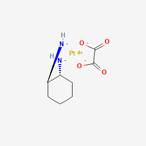
An organoplatinum complex in which the platinum atom is complexed with 1,2-diaminocyclohexane, and with an oxalate ligand which is displaced to yield active this compound derivatives. These derivatives form inter- and intra-strand DNA crosslinks that inhibit DNA replication and transcription. This compound is an antineoplastic agent that is often administered with FLUOROURACIL and FOLINIC ACID in the treatment of metastatic COLORECTAL NEOPLASMS.
Properties
Key on ui mechanism of action |
Oxaliplatin undergoes nonenzymatic conversion in physiologic solutions to active derivatives via displacement of the labile oxalate ligand. Several transient reactive species are formed, including monoaquo and diaquo DACH platinum, which covalently bind with macromolecules. Both inter and intrastrand Pt-DNA crosslinks are formed. Crosslinks are formed between the N7 positions of two adjacent guanines (GG), adjacent adenine-guanines (AG), and guanines separated by an intervening nucleotide (GNG). These crosslinks inhibit DNA replication and transcription. Cytotoxicity is cell-cycle nonspecific. |
|---|---|
CAS No. |
61825-94-3 |
Molecular Formula |
C8H14N2O4Pt |
Molecular Weight |
397.29 g/mol |
IUPAC Name |
[(1R,2R)-2-azanidylcyclohexyl]azanide;oxalic acid;platinum(2+) |
InChI |
InChI=1S/C6H12N2.C2H2O4.Pt/c7-5-3-1-2-4-6(5)8;3-1(4)2(5)6;/h5-8H,1-4H2;(H,3,4)(H,5,6);/q-2;;+2/t5-,6-;;/m1../s1 |
InChI Key |
DRMCATBEKSVAPL-BNTLRKBRSA-N |
SMILES |
C1CCC(C(C1)[NH-])[NH-].C(=O)(C(=O)[O-])[O-].[Pt+4] |
Isomeric SMILES |
C1CC[C@H]([C@@H](C1)[NH-])[NH-].C(=O)(C(=O)O)O.[Pt+2] |
Canonical SMILES |
C1CCC(C(C1)[NH-])[NH-].C(=O)(C(=O)O)O.[Pt+2] |
Appearance |
white solid powder |
boiling_point |
100ºC |
Other CAS No. |
63121-00-6 61825-94-3 |
physical_description |
Solid |
Pictograms |
Irritant; Health Hazard |
Purity |
>98% (or refer to the Certificate of Analysis) |
Related CAS |
63121-00-6 63121-00-6 (SP-4-2 (trans)) |
shelf_life |
>10 years if stored properly |
solubility |
Soluble in water at 4 mg/mL and DMSO at 20 mg/mL; slightly soluble in methanol; insoluble in ethanol. |
storage |
Dry, dark and at 0 - 4 C for short term (days to weeks) or -20 C for long term (months to years). |
Synonyms |
1,2 Diaminocyclohexane Platinum Oxalate 1,2-diaminocyclohexane platinum oxalate 1,2-diamminocyclohexane(trans-1)oxolatoplatinum(II) ACT 078 ACT-078 ACT078 cis-oxalato-(trans-l)-1,2-diaminocyclohexane-platinum(II) Eloxatin Eloxatine L-OHP cpd oxalato-(1,2-cyclohexanediamine)platinum II oxaliplatin oxaliplatin, (SP-4-2-(1R-trans))-isomer oxaliplatin, (SP-4-2-(1S-trans))-isomer oxaliplatin, (SP-4-3-(cis))-isomer oxaliplatine Platinum(2+) ethanedioate (1R,2R)-1,2-cyclohexanediamine (1:1:1) platinum(II)-1,2-cyclohexanediamine oxalate |
Origin of Product |
United States |
Molecular Mechanisms of Oxaliplatin Action
DNA Adduct Formation and Damage Response
Upon entering a cell, oxaliplatin undergoes a process called aquation, where the oxalate ligand is displaced by water molecules, making the platinum complex reactive. patsnap.com This activated form then binds to DNA bases, primarily forming adducts. patsnap.com The formation of these DNA adducts is central to this compound's cytotoxic effect. nih.gov
Intrastrand and Interstrand DNA Crosslinks
This compound forms different types of DNA crosslinks. The most prevalent are intrastrand crosslinks, which occur between two adjacent purine bases on the same DNA strand, predominantly between two guanine residues (GG) or, less frequently, between guanine and adenine (GA). aacrjournals.orgscirp.orgnih.govnih.gov this compound also forms interstrand crosslinks, which are covalent bonds between bases on opposite DNA strands, and DNA-protein crosslinks. aacrjournals.orgnih.govoup.com While interstrand crosslinks are considered highly toxic DNA lesions, they constitute a smaller fraction of the total adducts formed by this compound compared to intrastrand crosslinks. aacrjournals.orgscirp.orgnih.gov
Specific DNA Binding Sites (e.g., N7-Guanine)
The reactive platinum species of this compound primarily binds to the N7 position of guanine residues in DNA. patsnap.comscirp.orgnih.govnih.gov Binding to adenine bases at the N7 position also occurs, though less frequently than with guanine. patsnap.comscirp.orgmetu.edu.tr This binding forms monoadducts, which can then react further with an adjacent purine base to form the more stable and cytotoxic diadducts, resulting in intrastrand or interstrand crosslinks. nih.govnih.govresearchgate.net
Inhibition of DNA Replication and Transcription
The formation of platinum-DNA adducts, particularly the intrastrand and interstrand crosslinks, causes distortions in the DNA helix. scirp.org These structural alterations act as physical blocks, preventing the enzymes involved in DNA replication (DNA polymerases) and transcription (RNA polymerases) from moving along the DNA template. wikipedia.orgpatsnap.commedchemexpress.comaacrjournals.orgscirp.orgmdpi.com This inhibition of DNA replication and transcription is a key mechanism by which this compound hinders cell division and protein synthesis, ultimately leading to cell cycle arrest and cell death. patsnap.commedchemexpress.comscirp.org
Activation of Homologous Recombination Repair Pathways
The DNA damage induced by this compound, including DNA crosslinks and double-strand breaks, activates cellular DNA damage response pathways. patsnap.comamegroups.org One of the key repair mechanisms involved in addressing this compound-induced DNA damage, particularly interstrand crosslinks and double-strand breaks, is homologous recombination (HR) repair. patsnap.comoup.comamegroups.orgnih.govdovepress.com HR is a major pathway for repairing double-strand breaks and is crucial for maintaining genomic integrity. amegroups.orgdovepress.com Activation of HR can contribute to resistance to this compound in cancer cells. nih.gov
Cellular Pathway Modulation
Apoptosis Induction Mechanisms
This compound-induced DNA damage triggers signaling cascades that lead to the induction of apoptosis, a form of programmed cell death essential for eliminating damaged or unwanted cells. patsnap.commedchemexpress.comscirp.org Several mechanisms contribute to this compound-induced apoptosis. One important pathway involves the activation of the tumor suppressor protein p53, which can regulate the cell cycle and promote apoptosis in response to DNA damage. patsnap.commdpi.comnih.govmedwinpublishers.com this compound treatment has been shown to upregulate p53 in some cancer cell lines. medwinpublishers.com Additionally, this compound can activate the mitochondria-mediated apoptotic pathway, characterized by the release of cytochrome c from the mitochondria into the cytosol. patsnap.comnih.govnih.gov This release subsequently activates caspases, a family of proteases that execute the final stages of apoptosis, including caspase-3. patsnap.comnih.govnih.gov Studies have shown that this compound treatment results in caspase-3 activation. nih.gov The balance between pro-apoptotic and anti-apoptotic proteins, such as those in the Bcl-2 family, plays a critical role in regulating this compound-induced apoptosis. mdpi.comnih.govmedwinpublishers.com this compound may also induce apoptosis through mechanisms involving oxidative stress and the generation of reactive oxygen species (ROS). patsnap.commdpi.comijbs.com Increased ROS levels can cause further cellular damage and contribute to apoptosis. patsnap.comijbs.com Activation of signaling pathways like the p38 MAPK pathway has also been implicated as a key proapoptotic mediator of this compound-induced cell death in some cell lines. ijbs.comaacrjournals.org
Intrinsic Apoptotic Pathway Activation
The intrinsic apoptotic pathway, also known as the mitochondrial pathway, is a major route through which this compound exerts its cytotoxic effects. nih.govdiva-portal.org This pathway is initiated by intracellular stress signals, such as DNA damage induced by this compound, which converge on the mitochondria. spandidos-publications.com
This compound treatment can lead to mitochondrial dysfunction and derangement. nih.govresearchgate.net A key event in the activation of the intrinsic pathway is the mitochondrial outer membrane permeabilization (MOMP), which results in the release of pro-apoptotic factors from the intermembrane space into the cytosol. spandidos-publications.comjci.org Cytochrome C is a crucial protein released from the mitochondria during this process. nih.govspandidos-publications.comresearchgate.netunimib.itmdpi.com Once in the cytosol, cytochrome C binds with Apaf-1 and procaspase-9 to form the apoptosome, a molecular complex that activates caspase-9. jci.orgmdpi.com Activated caspase-9 subsequently cleaves and activates downstream effector caspases, such as caspase-3, leading to the execution of apoptosis. diva-portal.orgjci.orgmdpi.com Studies have shown that this compound can induce the cytosolic release of cytochrome C. nih.govresearchgate.net
The intrinsic apoptotic pathway is tightly regulated by the balance between pro-apoptotic and anti-apoptotic proteins, particularly those belonging to the Bcl-2 family. spandidos-publications.comnih.gov Anti-apoptotic proteins like Bcl-2 are typically localized on the outer mitochondrial membrane and prevent MOMP and the release of cytochrome C. spandidos-publications.comnih.gov this compound can influence the expression and activity of these proteins. Some research indicates that this compound treatment can lead to decreased expression of anti-apoptotic protein Bcl-2, thereby promoting the release of cytochrome C and activating the intrinsic pathway. nih.govoncotarget.comspandidos-publications.com Conversely, overexpression of anti-apoptotic Bcl-2 family members has been associated with resistance to chemotherapy, including this compound. oncotarget.com Bcl-2 interacts with various proteins, including pro-apoptotic members like Bax and Bak, and its binding to Bax can block the release of cytochrome C. mdpi.comnih.govmdpi.com
Extrinsic Apoptotic Pathway Activation
The extrinsic apoptotic pathway is initiated by the binding of extracellular death ligands to specific death receptors on the cell surface, leading to the formation of the death-inducing signaling complex (DISC). spandidos-publications.comjci.org While the intrinsic pathway is considered a primary mechanism for this compound-induced apoptosis, the extrinsic pathway also contributes to its cytotoxic effects in certain cell types. nih.govwjgnet.com
Activation of the extrinsic pathway involves the recruitment and activation of initiator caspases, particularly caspase-8, within the DISC. spandidos-publications.comjci.orgiiarjournals.org Activated caspase-8 can directly cleave and activate effector caspases or cleave the pro-apoptotic Bcl-2 family protein Bid. spandidos-publications.comjci.org Cleavage of Bid generates a truncated form (tBid) which translocates to the mitochondria and promotes MOMP, thereby linking the extrinsic and intrinsic pathways and amplifying the apoptotic signal. spandidos-publications.comjci.orgresearchgate.netplos.org Studies have shown that this compound can increase caspase-8 activity and Bid expression in certain cancer cell lines, indicating activation of the extrinsic pathway. nih.govwjgnet.com
The extrinsic pathway is initiated by the engagement of death receptors, such as Death Receptor 4 (DR4) and Death Receptor 5 (DR5), by their respective ligands (e.g., TRAIL). spandidos-publications.comjci.org While this compound's primary action is intracellular DNA damage, there is evidence suggesting its influence on death receptors. Some studies indicate that this compound, particularly in combination with other agents, can lead to increased expression or altered localization of death receptors like FasL and DR4, contributing to the activation of the extrinsic pathway. wjgnet.comiiarjournals.org The clustering of these receptors upon ligand binding is crucial for the formation of the DISC and subsequent caspase activation. jci.org
The induction of apoptosis by this compound is a multi-faceted process initiated by DNA damage. patsnap.com This damage serves as a signal that converges on the cellular apoptotic machinery, activating distinct yet interconnected pathways that culminate in controlled cell dismantling. nih.govspandidos-publications.com
Intrinsic Apoptotic Pathway Activation
The intrinsic apoptotic pathway, often referred to as the mitochondrial pathway, represents a significant route through which this compound mediates cell death. nih.govdiva-portal.org This pathway is triggered by intracellular stressors, including the DNA lesions induced by this compound, which impact mitochondrial integrity and function. spandidos-publications.com
A pivotal event in the activation of the intrinsic pathway is the permeabilization of the mitochondrial outer membrane (MOMP). spandidos-publications.comjci.org this compound can induce mitochondrial dysfunction, leading to MOMP and the subsequent release of pro-apoptotic factors from the mitochondrial intermembrane space into the cytosol. nih.govspandidos-publications.comresearchgate.netunimib.itmdpi.com Among these factors, Cytochrome C plays a critical role. nih.govspandidos-publications.comresearchgate.netunimib.itmdpi.com Upon its release into the cytosol, Cytochrome C interacts with Apaf-1 and procaspase-9, facilitating the formation of the apoptosome. jci.orgmdpi.com This complex serves as a platform for the autoactivation of caspase-9, an initiator caspase. jci.orgmdpi.com Activated caspase-9 then cleaves and activates executioner caspases, notably caspase-3, which are responsible for dismantling the cell. diva-portal.orgjci.orgmdpi.com Research indicates that this compound treatment can lead to the cytosolic translocation of Cytochrome C. nih.govresearchgate.net
The intrinsic apoptotic pathway is under stringent control by the balance between pro-apoptotic and anti-apoptotic proteins, particularly members of the Bcl-2 protein family. spandidos-publications.comnih.gov Anti-apoptotic proteins like Bcl-2 reside on the outer mitochondrial membrane and function to prevent MOMP and the release of pro-apoptotic factors like Cytochrome C. spandidos-publications.comnih.gov this compound can modulate the expression and activity of these regulatory proteins. Studies have demonstrated that this compound treatment can result in a decrease in the expression levels of the anti-apoptotic protein Bcl-2, thereby favoring the release of Cytochrome C and the activation of the intrinsic pathway. nih.govoncotarget.comspandidos-publications.com Conversely, the overexpression of anti-apoptotic Bcl-2 family proteins has been linked to reduced sensitivity and resistance to various chemotherapeutic agents, including this compound. oncotarget.com Bcl-2 interacts with numerous proteins, including pro-apoptotic counterparts such as Bax and Bak, and its binding to Bax is known to inhibit the release of Cytochrome C from mitochondria. mdpi.comnih.govmdpi.com
Mitochondrial Derangement and Cytochrome C Release
Extrinsic Apoptotic Pathway Activation
The extrinsic apoptotic pathway is triggered by the engagement of death receptors on the cell surface by their specific ligands, leading to the assembly of the death-inducing signaling complex (DISC). spandidos-publications.comjci.org While the intrinsic pathway is a major contributor to this compound-induced apoptosis, the extrinsic pathway also plays a role in mediating cell death in response to this drug in certain cellular contexts. nih.govwjgnet.com
Activation of the extrinsic pathway involves the recruitment and activation of initiator caspases, primarily caspase-8, within the DISC. spandidos-publications.comjci.orgiiarjournals.org Activated caspase-8 can directly activate executioner caspases or cleave the pro-apoptotic Bcl-2 family member Bid. spandidos-publications.comjci.org The cleavage of Bid generates a truncated form, tBid, which then translocates to the mitochondria. spandidos-publications.comjci.orgresearchgate.netplos.org tBid promotes MOMP, thereby establishing a link between the extrinsic and intrinsic pathways and amplifying the apoptotic signal. spandidos-publications.comjci.orgresearchgate.netplos.org Research indicates that this compound can enhance caspase-8 activity and increase Bid expression in certain cancer cell lines, suggesting the involvement of the extrinsic pathway. nih.govwjgnet.com
The extrinsic pathway is initiated by the interaction of death ligands (e.g., TRAIL) with their cognate death receptors, such as Death Receptor 4 (DR4) and Death Receptor 5 (DR5), on the cell membrane. spandidos-publications.comjci.org Although this compound's primary mechanism involves intracellular DNA damage, there is evidence suggesting its influence on death receptors. Some studies indicate that this compound, particularly when used in combination with other agents, can lead to the upregulation or altered localization of death receptors like FasL and DR4, contributing to the activation of the extrinsic apoptotic cascade. wjgnet.comiiarjournals.org The clustering of these receptors upon ligand binding is essential for the formation of the DISC and the subsequent activation of caspases. jci.org
Caspase-8 Activity and Bid Expression
Autophagy-Associated Apoptosis
Autophagy is a cellular process involving the degradation and recycling of damaged organelles and proteins. nih.gov While often acting as a survival mechanism in response to cellular stress, autophagy can also contribute to cell death or be intricately linked with apoptosis. nih.govmdpi.com Studies investigating the role of autophagy in this compound-treated cancer cells have yielded complex results, suggesting a context-dependent function.
In some instances, this compound treatment has been shown to induce autophagy in cancer cells. nih.gov This induction can be a protective response, helping cells cope with the stress imposed by the drug, and its inhibition may enhance this compound's cytotoxic effects. nih.govnih.govresearchgate.net For example, in Caco-2 colorectal cancer cells, this compound treatment increased the ratio of MAP1LC3B-II to Actin, a marker of autophagy, and inhibiting autophagy with pharmacological agents or siRNA targeting essential autophagy genes like ATG5 and Beclin1 potentiated this compound-induced cell death. nih.gov This suggests that in these cells, autophagy acts as a pro-survival mechanism against this compound. nih.gov
Conversely, other research indicates that this compound may reduce the expression of autophagy-related proteins like Atg5, beclin-1, Atg7, LC3-I, and LC3-II in a dose-dependent manner in certain cell lines, suggesting a potential inhibition of autophagy. nih.gov The interplay between this compound, autophagy, and apoptosis is a critical area of research, particularly in understanding mechanisms of drug resistance. mdpi.comresearchgate.net Autophagy-associated apoptosis, where autophagy directly contributes to cell death, has also been implicated as a mechanism facilitated by this compound in certain contexts. mdpi.com
Cell Cycle Perturbation and Arrest
The cell cycle is a tightly regulated series of events that leads to cell division. Anticancer agents often target specific phases of the cell cycle to inhibit cancer cell proliferation. This compound has been shown to induce cell cycle perturbation and arrest in various cancer cell lines. nih.govaacrjournals.org
A primary effect of this compound is the induction of G2/M cell cycle arrest, although a transient S phase delay has also been observed. nih.gov Studies using flow cytometry have demonstrated that this compound treatment leads to an accumulation of cells in the G2/M phase. nih.govaacrjournals.org For instance, in HT29, MCF7, Hela, and A549 cell lines, this compound treatment resulted in significant cell cycle modifications, with a prominent G2/M arrest. nih.gov The extent and reversibility of this arrest can vary depending on the cell line and treatment duration. nih.gov
Research has also explored the impact of this compound on cell cycle regulatory proteins. While not explicitly detailed in the provided snippets for this compound alone, studies involving combinations with this compound have shown alterations in the levels of cyclins A and B, as well as cdc2 and cdk2, which are key regulators of G2/M progression. tandfonline.com The induction of cell cycle arrest by this compound is considered a crucial mechanism contributing to its cytotoxic effects, preventing cancer cells from dividing and proliferating. researchgate.netmdpi.com
Reactive Oxygen Species Generation and Oxidative Stress Response
This compound treatment can lead to the generation of reactive oxygen species (ROS), which are highly reactive molecules that can cause oxidative stress and damage to cellular components, including DNA, lipids, and proteins. nih.govnih.govijbs.com Oxidative stress is recognized as an important factor in the toxicity of this compound, including mitochondrial toxicity. nih.gov
Studies have shown that this compound increases intracellular ROS levels in cancer cells. nih.govijbs.com This increased ROS production can contribute to this compound-induced DNA damage and apoptosis. nih.gov For example, in colorectal cancer cells, this compound treatment statistically significantly increased ROS levels. nih.gov The generation of ROS by this compound is considered a critical event in platinum-induced cell death. plos.org
The cellular response to this compound-induced oxidative stress involves various antioxidant defense mechanisms. However, excessive ROS generation can overwhelm these defenses, leading to cellular damage and the activation of cell death pathways. nih.govijbs.com The relationship between ROS, autophagy, and apoptosis in the context of this compound treatment is complex, with ROS potentially triggering autophagy and endoplasmic reticulum (ER) stress, which can in turn influence cell fate. nih.govplos.org
Immunogenic Cell Death Pathways
Immunogenic cell death (ICD) is a mode of cell death that elicits an adaptive immune response against dead-cell-associated antigens. This process involves the release of damage-associated molecular patterns (DAMPs) that can activate dendritic cells and prime T cell responses against cancer cells. researchgate.nettargetedonc.com this compound has been identified as an inducer of ICD. researchgate.nettargetedonc.comwjon.org
Compared to other platinum compounds like cisplatin and irinotecan, this compound has shown a greater capacity to induce ICD. targetedonc.comwjon.org This involves the translocation of calreticulin and other chaperones from the endoplasmic reticulum to the cell membrane, as well as the release of molecules like ATP and HMGB1. researchgate.net These events act as "eat-me" and "danger" signals, promoting the uptake of tumor antigens by dendritic cells and subsequent activation of cytotoxic T lymphocytes. researchgate.netresearchgate.net
Research suggests that the immunogenic properties of this compound contribute to its therapeutic efficacy, particularly in colorectal cancer. targetedonc.comwjon.org The activation of the host immune system by this compound-induced ICD can enhance the antitumor effect. researchgate.net Studies have explored the association between genetic variations in ICD pathways and the efficacy of this compound-based chemotherapy. targetedonc.com The cGAS-STING signaling pathway has also been implicated in this compound-induced ICD in certain cancer types. biorxiv.org
Nucleolar Stress Response
The nucleolus is a dynamic structure within the nucleus primarily involved in ribosome biogenesis. Disruptions to nucleolar function or ribosome biogenesis can trigger a nucleolar stress response, which can lead to cell cycle arrest or apoptosis, often via activation of the tumor suppressor protein p53. nih.govnih.govaacrjournals.org Emerging evidence indicates that this compound, unlike cisplatin or carboplatin, can induce cytotoxicity by specifically disrupting ribosome biogenesis and triggering nucleolar stress. nih.govnih.govbiorxiv.orgresearchgate.net
This compound causes early nucleolar disruption, characterized by the inhibition of ribosomal RNA (rRNA) synthesis and the relocalization of nucleolar proteins like nucleophosmin (NPM1). nih.govnih.govbiorxiv.org These effects can occur at concentrations below those required for significant DNA damage, suggesting that nucleolar stress is an important and potentially early event in this compound's mechanism of action. nih.govnih.gov
The induction of nucleolar stress by this compound appears to be linked to DNA damage signaling involving ATM and ATR kinases, although it does not necessarily involve substantial nucleolar DNA damage itself. biorxiv.org The nonlabile diaminocyclohexane (DACH) ligand of this compound has been identified as important for its ability to induce nucleolar stress. nih.gov Proteomic profiling of cells treated with this compound has revealed downregulation of ribosomal proteins, further supporting the notion of disrupted ribosome biogenesis and nucleolar stress as key responses. aacrjournals.org This nucleolar and ribosomal stress can subsequently trigger apoptosis through both p53-dependent and independent pathways. aacrjournals.org
Mechanisms of Oxaliplatin Resistance
DNA Damage Response and Repair Alterations
Tumor cells can develop resistance to oxaliplatin by altering their DNA damage response and repair pathways. These alterations can lead to enhanced tolerance to the platinum-DNA adducts formed by this compound, as well as more efficient mechanisms for repairing this damage.
Enhanced Tolerance to Platinum-DNA Adducts
Enhanced tolerance refers to the ability of resistant cells to endure the presence of platinum-DNA adducts without triggering the normal cell death pathways. While this compound forms DNA adducts that block replication and transcription, resistant cells can develop mechanisms to bypass these lesions or tolerate their presence, allowing for continued cell division. Studies have shown that resistant cell lines can exhibit increased tolerance to adducts in DNA. nih.gov This tolerance can range from 3.1 to 7.6-fold in some resistant sub-lines. nih.gov This suggests that even if adducts are formed, the cellular machinery in resistant cells is less sensitive to their cytotoxic effects.
Upgraded Excision Repair Mechanisms (e.g., ERCC1 pathway)
The nucleotide excision repair (NER) pathway is a major DNA repair mechanism involved in removing bulky DNA lesions, including those formed by platinum compounds like this compound. aacrjournals.orgiiarjournals.org The excision repair cross-complementing group 1 (ERCC1) protein, in complex with XPF, plays a critical role in the NER pathway by creating an incision 5' to the damaged site. iiarjournals.orgecancer.orgiiarjournals.orgmdpi.com
Increased activity or expression of the NER pathway, particularly involving ERCC1, has been strongly implicated in this compound resistance. iiarjournals.orgecancer.orgiiarjournals.orgmdpi.com Studies have demonstrated that this compound treatment can induce ERCC1 gene expression in resistant, but not sensitive, colorectal cancer cell lines. iiarjournals.orgiiarjournals.org This induction of ERCC1 mRNA and protein in resistant cells suggests that the NER pathway is upregulated in response to this compound-induced DNA damage, leading to more efficient repair and thus resistance. iiarjournals.orgiiarjournals.org
The level of ERCC1 expression has been shown to correlate with the effectiveness of this compound treatment in patients with colorectal cancer. ecancer.org High levels of ERCC1 expression are associated with reduced sensitivity to this compound. ecancer.orgmdpi.com
Furthermore, studies using siRNA-mediated silencing of ERCC1 have shown that reducing ERCC1 levels in resistant cell lines can restore sensitivity to this compound, leading to increased apoptosis. iiarjournals.orgiiarjournals.org This provides strong evidence for the direct role of ERCC1 in mediating this compound resistance.
Replication Bypass Mechanisms
Replication bypass, also known as translesion synthesis (TLS), is a mechanism where specialized DNA polymerases can replicate past DNA lesions that would normally stall the high-fidelity replicative polymerases. This process can contribute to drug resistance by allowing cells with damaged DNA to continue dividing.
Studies suggest that replication bypass mechanisms can discriminate between cisplatin and this compound adducts. aacrjournals.org Human DNA polymerases such as pol β, pol γ, and pol η have been shown to replicate past this compound-GG adducts more efficiently than cisplatin-GG adducts in vitro. aacrjournals.orgnih.govaacrjournals.org The bulky diaminocyclohexane (DACH) ligand of this compound may influence how these polymerases interact with the adducts, potentially facilitating bypass. nih.govpatsnap.com Overexpression of certain polymerases involved in bypass has been linked to this compound resistance in some cancer cell lines. aacrjournals.org
Cellular Drug Accumulation and Efflux Dynamics
The amount of this compound that reaches its intracellular target (DNA) is a critical determinant of its efficacy. Alterations in the mechanisms that control the influx and efflux of the drug across the cell membrane can significantly impact intracellular drug accumulation and contribute to resistance.
Decreased Cellular Uptake Mechanisms
Reduced cellular uptake of this compound is a documented mechanism of resistance. nih.govmdpi.comaacrjournals.orgresearchgate.netnih.govnih.gov Studies have shown that this compound-resistant cell lines exhibit significantly lower intracellular accumulation of the drug compared to sensitive cell lines. nih.govaacrjournals.orgnih.govmdpi.com This decreased uptake can be a significant factor, sometimes accounting for a substantial reduction in intracellular platinum levels. nih.gov
Several transporters are involved in the cellular uptake of platinum compounds, including organic cation transporters (OCTs) and the human copper transporter 1 (hCTR1). mdpi.comresearchgate.net Changes in the expression levels or function of these transporters can lead to decreased this compound influx. For instance, reduced expression of OCT1-3 has been correlated with decreased cellular this compound accumulation and resistance in colorectal cancer cell lines. mdpi.comnih.gov
Increased Cellular Efflux Mechanisms
In addition to decreased uptake, increased efflux of this compound from the cell can also contribute to lower intracellular drug concentrations and thus resistance. Efflux transporters, particularly ATP-binding cassette (ABC) transporters, can actively pump platinum compounds out of the cell. mdpi.comresearchgate.net
ATPases like ATP7A and ATP7B have been implicated in the intracellular trafficking and efflux of platinum drugs, including this compound. mdpi.comresearchgate.netnih.gov Increased expression of ATP7A has been shown to mediate resistance to this compound in ovarian cancer cells. aacrjournals.org This increased expression can lead to increased sequestration of platinum into vesicular fractions and reduced net accumulation in the cytoplasm and nucleus. aacrjournals.org
Another efflux transporter, ABCC10 (MRP7), has also been identified as a mediator of this compound efflux. nih.gov Higher expression of ABCC10 in colorectal cancer patients is associated with a greater likelihood of relapse and metastasis. nih.gov Knockdown of ABCC10 in resistant colorectal cancer cells increases intracellular this compound accumulation and enhances sensitivity to the drug, indicating that ABCC10-mediated efflux is a mechanism of resistance. nih.gov
Drug Inactivation Processes (e.g., Glutathione Binding, Metallothionein Segregation)
One of the key mechanisms by which cancer cells develop resistance to this compound is through drug inactivation. This can occur via the binding of this compound to intracellular molecules like glutathione (GSH) and metallothioneins (MTs). Once this compound enters the cytoplasm, it can undergo hydration, which facilitates its reaction with thiol-containing molecules such as GSH or metallothioneins. aacrjournals.orgresearchgate.net Binding to these molecules can effectively sequester the drug, preventing it from reaching its DNA target and forming cytotoxic adducts. researchgate.netnih.gov This decreased intracellular concentration of active platinum compounds contributes to reduced drug efficacy and the development of resistance. aacrjournals.orgdergipark.org.tr Studies have reported conflicting results regarding the precise association between the levels or activity of GSH and this compound resistance in vitro. aacrjournals.orgresearchgate.net
Role of Organic Cation Transporters (OCTs) and ABC Transporters
Cellular accumulation of this compound is a critical determinant of its efficacy, and alterations in drug transport proteins play a significant role in resistance. This compound uptake can be mediated by organic cation transporters (OCTs), specifically OCT1, OCT2, and OCT3 (SLC22A1-3). nih.govaacrjournals.org Studies have shown that the expression levels of OCTs can positively correlate with this compound absorption and sensitivity. rsc.org Conversely, decreased expression or function of these uptake transporters can lead to reduced intracellular drug concentrations and contribute to resistance. researchgate.net For instance, the transporter protein OCT-1 has been found to be present in this compound-sensitive colon cancer cells but absent in drug-resistant cells. researchgate.net
Efflux transporters, particularly those belonging to the ATP-binding cassette (ABC) transporter superfamily, are also implicated in this compound resistance. aacrjournals.orgrsc.org These transporters can actively pump drugs out of the cell, thereby lowering intracellular drug accumulation. rsc.org The ABCC family of transporters, also known as multidrug resistance proteins (MRPs), are involved in the glutathione (GSH)-mediated export of platinum drugs, including this compound. aacrjournals.orgresearchgate.net Overexpression of certain ABC transporters, such as ABCC2, ABCC3, and ABCG2, has been demonstrated to be beneficial for the efficacy of this compound in some cell lines, suggesting their role in transport-mediated resistance. oup.comresearchgate.net However, the association between the expression of certain ABC transporters like ABCB1 (MDR1) and this compound resistance has shown inconsistent results, highlighting the need for further investigation. researchgate.net
Cell Death Pathway Dysregulation
This compound induces cell death primarily through the induction of apoptosis following DNA damage. Resistance to this compound can arise from dysregulation of cell death pathways, allowing cancer cells to evade the cytotoxic effects of the drug. elifesciences.orgresearchgate.net
Resistance to Apoptosis
Resistance to this compound-induced apoptosis is a significant mechanism of acquired resistance. elifesciences.orgresearchgate.net This can involve alterations in the expression or function of key proteins that regulate the apoptotic cascade. Proteins involved in the intrinsic apoptotic pathway, such as members of the Bcl-2 family and the tumor suppressor p53, are often deregulated in drug-resistant cancers. mdpi.com For example, studies have shown that activated AKT can directly influence apoptotic pathway regulators like Bcl-2 family members, and PI3K/AKT signaling can increase Bcl-2 expression and activity, promoting cell survival and reducing chemotherapy-induced apoptosis. mdpi.com Alterations in the balance between pro-apoptotic (e.g., Bax, BBC3/PUMA) and anti-apoptotic (e.g., Bcl-2, Bcl-XL) proteins can contribute to an impaired apoptotic response. nih.govresearchgate.net Loss of Bax expression due to mutation has been observed in some this compound-resistant cell lines, suggesting a role for altered mitochondrial-mediated apoptosis in resistance. researchgate.net Impairment of the extrinsic apoptotic pathway has also been implicated, with overexpression of proteins like MMP7, which can degrade FasL, preventing apoptosis progression. researchgate.net
Autophagy Modulation in Resistance
Autophagy, a cellular process involving the degradation and recycling of damaged organelles and proteins, can play a dual role in cancer, sometimes promoting cell survival and sometimes cell death. In the context of this compound resistance, autophagy is often upregulated in tumor cells in response to chemotherapy and can contribute to resistance. researchgate.netnih.gov this compound has been shown to modulate autophagy, leading to the development of resistance. mdpi.com For instance, this compound can trigger AMPK signaling and inhibit mTOR signaling to induce autophagy. mdpi.com Studies in this compound-resistant gastric cancer cells have shown increased autophagy, suggesting it may be a key factor in resistance development. mdpi.com Autophagic cell death, which includes autophagy-associated apoptosis, can facilitate cellular resistance to platinum therapy and drug resistance. mdpi.com Inhibition of autophagy has been shown in some research to reverse or overcome this compound resistance. nih.govnih.gov Mechanical stress in the tumor microenvironment can also trigger autophagy activation, which is correlated with increased resistance to this compound treatment. nih.gov
Signaling Pathway Aberrations
Aberrant activation of various signaling pathways can promote cancer cell survival, proliferation, and resistance to chemotherapy, including this compound.
PI3K/AKT Signaling Pathway Activation
Hyperactivation of the phosphoinositide 3-kinase (PI3K)/AKT signaling pathway is frequently linked to this compound resistance. mdpi.commdpi.comnih.gov This pathway is a key regulator of cell survival, proliferation, and apoptosis. nih.govspandidos-publications.com Activated AKT, phosphorylated by PI3K, can directly influence apoptotic regulators, increasing anti-apoptotic protein activity and promoting cell survival, thereby lowering chemotherapy-induced apoptosis. mdpi.comnih.gov Studies have demonstrated increased constitutive activity of the PI3K/AKT pathway in cancer cells with acquired resistance to this compound. dovepress.com Inhibition of the PI3K/AKT pathway has been shown to increase the sensitivity of cancer cells to this compound. nih.govspandidos-publications.com For example, studies in cholangiocarcinoma and liver cancer cells have shown that inhibiting PI3K or its downstream target mTOR can enhance this compound efficacy. nih.govspandidos-publications.com The downregulation of PTEN, a negative regulator of the PI3K/AKT pathway, has also been observed in this compound-resistant cells, contributing to increased PI3K/AKT phosphorylation and reduced response to this compound-induced cell death. nih.gov
Table 1: and Associated Factors
| Mechanism | Associated Factors |
| Drug Inactivation Processes | Glutathione (GSH) binding aacrjournals.orgresearchgate.netmdpi.comnih.gov, Metallothionein (MT) segregation aacrjournals.orgresearchgate.netmdpi.comnih.gov |
| Transporter Alterations | Decreased uptake by OCT1, OCT2, OCT3 (SLC22A1-3) nih.govaacrjournals.orgresearchgate.net, Increased efflux by ABC transporters (e.g., ABCC family, ABCG2) rsc.orgoup.comresearchgate.net |
| Cell Death Dysregulation | Resistance to Apoptosis (e.g., altered Bcl-2 family, p53, Bax, IAPs) nih.govelifesciences.orgresearchgate.netmdpi.com, Autophagy modulation (often upregulation) researchgate.netmdpi.comnih.govnih.govnih.govtandfonline.com |
| Signaling Pathway Aberrations | Hyperactivation of PI3K/AKT pathway mdpi.commdpi.comnih.govspandidos-publications.comnih.govdovepress.com |
Table 2: Research Findings on PI3K/AKT Signaling in this compound Resistance
| Study Focus | Key Findings | Citations |
| Cholangiocarcinoma cells | Inhibition of PI3K/mTOR increased this compound cytotoxicity. Activated PI3K/AKT protected cells from this compound. | nih.gov |
| Liver cancer cells (HepG2R) | PI3K/AKT pathway stably activated in resistant cells. Inhibition of PI3K/AKT increased this compound sensitivity. | spandidos-publications.com |
| Colorectal cancer cells (HCT116) | Upregulation of PI3K/Akt pathway components in resistant cells. | aacrjournals.org |
| Colorectal cancer cells (SW480/R, HT29/R) | Downregulation of PTEN led to increased PI3K/AKT phosphorylation and reduced this compound sensitivity. | nih.gov |
| Colorectal cancer cells (HCT-8/L-OHP) | Activated PI3K/AKT pathway involved in resistance. Knockdown of KLK11 reversed resistance via PI3K/AKT suppression. | dovepress.com |
NF-κB Signaling Alterations
The NF-κB signaling pathway plays a crucial role in regulating numerous cellular functions, including those that contribute to tumor drug resistance. wjgnet.com Activation of the NF-κB pathway can enhance the expression of anti-apoptotic proteins, thereby making cancer cells more resistant to chemotherapy-induced apoptosis. researchgate.net Studies have shown that chemotherapy-induced activation of NF-κB signaling contributes to heightened drug resistance in tumor cells. wjgnet.com For instance, in this compound-resistant colorectal cancer cells, upregulation of NF-κB phosphorylation has been observed, leading to enhanced expression of ABCG2, a transporter protein, and subsequent attenuation of endoplasmic reticulum stress and inhibition of apoptosis. researchgate.net The NF-κB pathway can also interact with other signaling cascades, such as the PI3K-AKT pathway, forming extensive regulatory networks that influence tumor cell behavior, including growth, resistance to apoptosis, and migration. wjgnet.com PIPKIγ has been identified as a critical gene related to this compound resistance in colorectal cancer, promoting the expression of PD-L1 via activation of the NF-κB signaling pathway. thno.org Knockdown of PIPKIγ significantly down-regulated the phosphorylated level of P65, a component of NF-κB, while overexpression increased it, consistently affecting NF-κB transcriptional activity. thno.org
Epigenetic Modifications in Resistance Acquisition
Epigenetic modifications, which involve heritable changes in gene expression without alterations to the underlying DNA sequence, play a significant role in the acquisition of this compound resistance. wjgnet.com These modifications can deregulate genes involved in crucial cellular processes such as cell cycle regulation, apoptosis, DNA damage response, and DNA repair pathways. mdpi.com
Epigenetic silencing, a major mechanism causing the loss of tumor suppressor activity, is often caused by the integration of promoter hypermethylation and histone deacetylation. biomolther.org These processes are controlled by regulatory proteins such as DNA methyltransferases (DNMTs) and histone deacetylases (HDACs). biomolther.org Research indicates that gene methylation status is associated with chemotherapy response; for example, high methylation of the SRBC gene may lead to increased resistance to this compound in colorectal cancer cells. researchgate.net Promoter hypermethylation of CAVIN3 has also been associated with resistance to this compound in colorectal cancer cell lines and predicts shorter progression-free survival in patients with metastatic disease. researchgate.net In renal cell carcinoma, loss of the organic cation transporter OCT2, a main factor contributing to this compound resistance, is linked to DNA hypermethylation and histone methylation at the OCT2 promoter. nih.gov Histone acetylation also regulates OCT2 repression in these cells. nih.gov Combined treatment with a DNA methylation inhibitor (decitabine) and a histone deacetylase inhibitor (vorinostat) significantly increased OCT2 expression in renal cell carcinoma cell lines, sensitizing them to this compound. nih.gov
The high-order three-dimensional (3D) conformation of the genome can be altered during the acquisition of this compound resistance. nih.gov Studies investigating chromatin conformation alterations in this compound-resistant colorectal cancer cells using techniques like DLO Hi-C, ChIP-seq, and RNA-seq have revealed significant changes. nih.gov Results indicate that a substantial percentage of genome regions undergo A/B compartment transformation after drug resistance develops. nih.gov Analysis of genes in these converted compartments suggests that this compound resistance is related to a reduction in reactive oxygen species and enhanced metastatic capacity. nih.gov Furthermore, 3D structure mapping of DNA damage shows characteristic differences in the nuclear distribution of this compound-induced DNA damage between drug-resistant and sensitive cells. oup.com Damage increases in gene-poor chromatin at the nuclear periphery while decreasing in gene-rich regions located at nuclear speckles, suggesting a strategic redistribution of platinum drug-induced damage during chemoresistance development. oup.com
Changes in gene expression profiles, including those of long non-coding RNAs (lncRNAs), are implicated in this compound resistance. LncRNAs are important regulators of gene expression and can play key roles in drug function regulation and chemoresistance. spandidos-publications.com HOTAIR (HOX transcript antisense RNA) is one such lncRNA that has been investigated in the context of this compound resistance. nih.gov Studies have shown that HOTAIR expression is increased in hypoxia-treated colorectal cancer cell lines and tumors, and its knockdown sensitizes these cells to this compound. nih.govresearchgate.net High levels of HOTAIR have been associated with poor prognosis in colorectal cancer patients. spandidos-publications.com HOTAIR can contribute to resistance by acting as a competitive endogenous RNA (ceRNA) for miRNAs, thereby regulating the expression of target genes. nih.gov Besides HOTAIR, other lncRNAs such as CRNDE, MALAT1, ANRIL, GIHCG, CASC15, and MEG3 have also been identified as critical regulators of this compound resistance through various mechanisms, including sponging miRNAs and regulating target gene expression. mdpi.com For example, GIHCG is over-expressed in colorectal cancer and contributes to resistance to 5-fluorouracil and this compound. mdpi.com CASC15 contributes to this compound resistance by positively regulating the expression levels of ABCC1 by sponging miR-145. mdpi.com MEG3 down-regulation is associated with this compound resistance through the regulation of the miR-141/PDCD4 complex. mdpi.com
Deregulation of genes involved in cell cycle control and DNA repair pathways is a significant factor in this compound resistance. mdpi.com this compound forms DNA adducts, which ideally lead to cell cycle arrest and apoptosis in sensitive cells. mdpi.com However, specific genetic or epigenetic modifications can inhibit pathways like the p53-p21 pathway, leading to unchecked cell cycle progression and resistance. mdpi.com Increased capacity for DNA damage repair is a key mechanism underlying this compound resistance. researchgate.net The nucleotide excision repair (NER) system is involved in the repair of this compound-DNA adducts and has been shown to have predictive value for colorectal cancer treatment. nih.gov Alterations in DNA repair genes, such as ERCC1 and XRCC1, may also play a role. aacrjournals.org While decreased ERCC1 expression has been demonstrated in this compound-resistant cells, its correlation appears to be more with cell cycle response than direct DNA repair after platinum treatment. nih.gov Increased expression of genes involved in homologous recombination repair, such as RAD51 and Fanconi anemia genes, suggests that this pathway can contribute to this compound resistance. nih.gov Activation of DNA damage response kinases like CHK2 has also been implicated, with elevated levels of phosphorylated CHK2 observed in this compound-resistant colorectal cancer cells and tumor samples. nih.gov This activation contributes to accelerated DNA damage repair. nih.gov Aberrant activation of cell cycle regulation pathways correlates with resistance, and inhibiting components like CDK4/6 can enhance the antitumor effect of this compound by epigenetically suppressing DNA repair genes. pnas.org
Gene Expression Profile Changes (e.g., lncRNA HOTAIR)
Tumor Microenvironment Factors
The tumor microenvironment (TME) plays a crucial role in influencing therapeutic response and contributing to this compound resistance. wjgnet.comresearchgate.net The crosstalk between tumor cells and immune cells within the TME is particularly important. researchgate.net Tumor-associated macrophages (TAMs) are abundant in the TME and are implicated in platinum-based drug resistance in several cancers, including colorectal cancer. researchgate.net The density of tumor-infiltrating macrophages is significantly higher in this compound-resistant patients compared to sensitive patients. researchgate.net Macrophages can promote this compound resistance through various mechanisms, including the activation of autophagy in cancer cells. mdpi.comresearchgate.net Recent studies suggest that M2 TAMs can increase this compound resistance by enhancing METTL3-mediated m6A modification in cancer cells. researchgate.net The immunosuppressive environment created by the imbalance between immunosuppressive cells (like TAMs) and anti-tumor effector cells also contributes to therapeutic resistance. frontiersin.org
Table 1: Key Genes and Molecules Involved in this compound Resistance Mechanisms
| Mechanism | Gene/Molecule | Role in Resistance | Relevant Section |
| NF-κB Signaling Alterations | NF-κB | Promotes anti-apoptotic proteins, enhances drug resistance. | 2.4.2 |
| PIPKIγ | Activates NF-κB, promotes PD-L1 expression. | 2.4.2 | |
| ABCG2 | Upregulated by NF-κB, reduces intracellular drug accumulation. | 2.4.2 | |
| Epigenetic Modifications | SRBC | Hypermethylation associated with increased resistance. | 2.5.1 |
| CAVIN3 | Promoter hypermethylation associated with resistance and poor prognosis. | 2.5.1 | |
| OCT2 | Transcriptional repression via DNA hypermethylation and histone modifications. | 2.5.1 | |
| HDACs, DNMTs | Enzymes involved in histone deacetylation and DNA methylation, targeted by inhibitors to restore sensitivity. | 2.5.1 | |
| HOTAIR (lncRNA) | Upregulated in resistant cells, sensitizes cells to this compound when knocked down. | 2.5.3 | |
| CRNDE, MALAT1, ANRIL, GIHCG, CASC15, MEG3 (lncRNAs) | Regulate resistance through various mechanisms (e.g., sponging miRNAs). | 2.5.3 | |
| Deregulation of Cell Cycle and DNA Repair | p53-p21 pathway | Inhibition leads to unchecked cell cycle progression and resistance. | 2.5.4 |
| ERCC1, XRCC1 | DNA repair genes potentially involved in resistance. | 2.5.4 | |
| RAD51, Fanconi genes | Involved in homologous recombination repair, contribute to resistance. | 2.5.4 | |
| CHK2 | Activation contributes to accelerated DNA damage repair. | 2.5.4 | |
| Tumor Microenvironment Factors | Tumor-Associated Macrophages (TAMs) | Abundant in TME of resistant patients, promote resistance via autophagy and m6A modification. | 2.6 |
| METTL3 | Involved in m6A modification mediated by TAMs, promotes resistance. | 2.6 |
Hypoxic Conditions and Chemoresistance
The tumor microenvironment, particularly the presence of hypoxic (low oxygen) conditions, plays a significant role in the development of chemoresistance to various agents, including this compound. Hypoxia is a common feature of solid tumors and is often associated with poor prognosis and reduced sensitivity to both chemotherapy and radiotherapy mdpi.comcapes.gov.brnih.gov. Cancer cells that adapt to hypoxic environments can develop resistance to this compound through a variety of mechanisms spandidos-publications.comresearchgate.net.
Detailed research findings highlight several pathways by which hypoxia contributes to this compound resistance:
Hypoxia-Inducible Factors (HIFs): HIFs, particularly HIF-1α, are key regulators of the cellular response to hypoxia mdpi.comresearchgate.net. Under hypoxic conditions, HIF-1α is stabilized and promotes the transcription of numerous genes involved in processes such as cell metabolism, proliferation, angiogenesis, and drug resistance researchgate.netnih.gov. HIF-1α has been shown to induce this compound resistance in colorectal cancer (CRC) cells researchgate.netnih.gov. This can occur through various mechanisms, including the upregulation of ATP-Binding Cassette (ABC) transporters like ABCB1 (MDR1), ABCC1 (MRP1), and ABCG2 (BCRP), which actively pump chemotherapeutic drugs out of the cells, thereby reducing intracellular drug concentration mdpi.comzlfzyj.comdovepress.com. Studies in hepatic carcinoma cells have also demonstrated that hypoxia-induced upregulation of HIF-1α correlates with increased expression of MDR1, MRP1, and BCRP1, contributing to this compound resistance zlfzyj.com.
Modulation of Apoptosis: Hypoxia can enhance the anti-apoptotic potential of cancer cells, making them less susceptible to this compound-induced cell death mdpi.com. HIF-1 can induce the expression of anti-apoptotic proteins like Survivin and suppress pro-apoptotic proteins like Caspase-9 mdpi.com. Additionally, hypoxia can decrease the level of apoptosis induced by this compound in CRC cells nih.gov.
Signaling Pathways: Hypoxia can activate various signaling pathways that promote drug resistance. For instance, HIF is known to cause this compound resistance in colorectal cancer by inducing miR-338-5p expression, which promotes IL-6 signaling. This, in turn, suppresses apoptosis via the STAT3/BCL2 pathway mdpi.comnih.gov. The IL-6/STAT3/Bcl2 pathway has been identified as a critical mechanism of hypoxia-induced drug resistance in CRC cells nih.gov. Studies have also investigated the role of JNK signaling; hypoxia-induced JNK activation was associated with resistance to this compound in colon cancer cell lines aacrjournals.org.
Non-Coding RNAs: Long non-coding RNAs (lncRNAs) have been implicated in hypoxia-mediated this compound resistance. For example, studies have shown that hypoxia increases the expression of HOX transcript antisense RNA (HOTAIR) in CRC cells, and HOTAIR contributes to this compound resistance by modulating the expression of zinc finger E-box binding homeobox 1 (ZEB1) through negative regulation of miR-1277-5p researchgate.netresearchgate.net. MicroRNAs (miRNAs) also play a role; research suggests that under hypoxic conditions, P53 may induce this compound resistance through the activation of miR-23a spandidos-publications.com.
Drug Penetration: While biological mechanisms are significant, impaired drug penetration into hypoxic regions of tumors due to the distance from blood vessels also contributes to reduced this compound effectiveness mdpi.comnih.govdovepress.com. Studies using three-dimensional spheroids have shown that a sensitivity gradient exists, with hypoxic regions being less sensitive to this compound, which correlates with lower levels of platinum-bound DNA adducts nih.govnih.gov. However, the resistance observed in hypoxic sub-populations often exceeds what can be explained by reduced drug penetrance alone, indicating the involvement of other cellular adaptive responses nih.govnih.gov.
Data from various studies illustrate the impact of hypoxia on this compound sensitivity in different cancer cell lines. For example, in HCT116 colorectal cancer cells, the IC50 (the concentration of drug required for 50% inhibition of cell growth) for this compound is significantly higher under hypoxic conditions compared to normoxic conditions nih.gov.
Here is a table summarizing representative data on this compound sensitivity under normoxic and hypoxic conditions:
| Cell Line | Condition | This compound IC50 (µM) | Fold Resistance (Hypoxia/Normoxia) | Source |
| HCT116 | Normoxia | 5.2 | - | nih.gov |
| HCT116 | Hypoxia | 10.1 | 1.9 | nih.gov |
| HCT116 | Normoxia | 2.4 (Clonogenic Assay) | - | nih.gov |
| HCT116 | Hypoxia | 5.9 (Clonogenic Assay) | 2.4 | nih.gov |
| HCT116 | Normoxia | ~10 | - | nih.gov |
| HCT116 | Hypoxia | ~25 | ~2.5 | nih.gov |
| HCT8 | Normoxia | ~10 | - | nih.gov |
| HCT8 | Hypoxia | ~20 | ~2.0 | nih.gov |
| HT29 | Normoxia | ~15 | - | nih.gov |
| HT29 | Hypoxia | ~25 | ~1.7 | nih.gov |
| HCT116 | Normoxia | 20 µM (Growth Inhibition) | - | spandidos-publications.com |
| HCT116 | Hypoxia | 20 µM (Growth Inhibition) | Reduced Sensitivity | spandidos-publications.com |
| HT29 | Normoxia | 20 µM (Growth Inhibition) | - | spandidos-publications.com |
| HT29 | Hypoxia | 20 µM (Growth Inhibition) | Reduced Sensitivity | spandidos-publications.com |
Note: IC50 values can vary depending on the assay method and experimental conditions used in different studies.
The data consistently demonstrate that hypoxic conditions lead to reduced sensitivity or increased resistance to this compound in various cancer cell lines, particularly those of colorectal origin nih.govspandidos-publications.comnih.govresearchgate.net. This underscores the importance of understanding and targeting hypoxia-induced resistance mechanisms to improve the efficacy of this compound-based chemotherapy.
Biomarkers in Oxaliplatin Response and Resistance
Predictive Biomarkers of Therapeutic Efficacy
Predictive biomarkers aim to identify patients who are most likely to benefit from oxaliplatin-based chemotherapy. Several potential biomarkers are under investigation, focusing on different aspects of this compound's mechanism of action and cellular processing.
This compound-DNA Adduct Levels in Peripheral Blood Mononuclear Cells (PBMCs)
This compound exerts its cytotoxic effects through the formation of covalent drug-DNA adducts. The level of these adducts in tumor cells is hypothesized to correlate with treatment response. However, obtaining tumor biopsies can be challenging. Research suggests that this compound-DNA adduct levels in peripheral blood mononuclear cells (PBMCs) may serve as a surrogate biomarker for tumor DNA adducts and predict response to this compound-based regimens like FOLFOX (a combination of leucovorin calcium, fluorouracil, and this compound). nih.govnih.govosti.govaacrjournals.orgescholarship.orgresearchgate.net
Studies have investigated the relationship between this compound-DNA adduct levels in PBMCs and tumor response in colorectal cancer patients. One pilot clinical study involving six colorectal cancer patients treated with FOLFOX chemotherapy found a significant correlation between this compound-DNA adduct levels in PBMCs and the mean tumor volume change of evaluable target lesions. nih.govnih.govosti.govaacrjournals.org This study utilized a diagnostic microdosing approach with 14C-labeled this compound and accelerator mass spectrometry (AMS) for sensitive quantification of adduct levels. nih.govosti.govaacrjournals.org Adduct levels induced by sub-therapeutic microdoses were found to be linearly proportional to those induced by therapeutic doses in both cell culture experiments and patient samples, suggesting the potential utility of diagnostic microdosing for predicting therapeutic adduct levels and treatment response. nih.govosti.govaacrjournals.org
Data from this pilot study indicated that PBMC adduct levels significantly correlated with tumor shrinkage (microdose: R2 = 0.82, p = 0.033; therapeutic dose: R2 = 0.65, p = 0.099). aacrjournals.org This supports the hypothesis that this compound-DNA adduct levels in PBMCs are proportional to tumor shrinkage caused by FOLFOX therapy and can potentially predict response. nih.govnih.govosti.gov
Oxidative Phosphorylation Activity
Oxidative phosphorylation (OXPHOS) is the primary method of ATP synthesis in aerobic conditions. Recent research suggests that the activity of oxidative phosphorylation pathways may serve as a predictive biomarker for this compound response in colorectal cancer. Studies analyzing patient samples and this compound-resistant and -sensitive cell lines have identified oxidative phosphorylation pathways as potentially valuable predictive biomarkers. researchgate.netnih.govresearchgate.netmdpi.comthno.org
Research using datasets from patients treated with this compound has indicated that non-responding patients tend to exhibit low oxidative phosphorylation activity. researchgate.netnih.govmdpi.com This suggests that low OXPHOS activity may be associated with a poor prognosis in the context of this compound treatment. researchgate.netnih.gov Analysis of differential gene expression between this compound non-responder and responder colorectal cancer patients has highlighted OXPHOS KEGG pathways as having predictive value, with a high area under the curve (AUC = 0.843). researchgate.netnih.govresearchgate.netmdpi.com
Machine Learning-Derived Molecular Signatures (e.g., FOLFOXai)
Prognostic Biomarkers in this compound Treatment
Genetic biomarkers, particularly those within DNA repair pathways, have shown promise as prognostic markers in this compound-based chemotherapy. frontiersin.orgresearchgate.net this compound's mechanism of action involves DNA damage, and the efficiency of DNA repair mechanisms can influence treatment outcome.
A systematic review identified 26 genetic biomarkers within 14 genes significantly associated with treatment outcome in this compound-based chemotherapy for colorectal cancer. frontiersin.orgresearchgate.net Promising genetic biomarkers included single nucleotide polymorphisms (SNPs) in genes such as ERCC1 (rs11615), XPC (rs1043953), XPD (rs13181), and XPG (rs17655), which are involved in the nucleotide excision repair (NER) pathway. frontiersin.orgresearchgate.net Other identified markers included SNPs in MNAT, MMR status, ATM protein expression, HIC1 tandem repeat D17S5, and PIN1 (rs2233678). frontiersin.orgresearchgate.net While these markers show promise, further prospective research, especially in specific patient populations like those with colorectal peritoneal metastases treated with cytoreductive surgery and hyperthermic intraperitoneal chemotherapy (CRS + HIPEC) with this compound, is needed for comprehensive validation. frontiersin.orgresearchgate.net
Biomarkers for this compound-Induced Peripheral Neurotoxicity (OXAIPN)
This compound-induced peripheral neurotoxicity (OXAIPN) is a major dose-limiting toxicity that can significantly impact the quality of life for patients and often necessitates dose reduction or treatment discontinuation. nih.govdntb.gov.uaresearchgate.netnih.govbiorxiv.orgmdpi.com Identifying biomarkers that can predict the risk and severity of OXAIPN is crucial for personalized treatment approaches and strategies to mitigate neurotoxicity.
Genetic Profiles (e.g., Single Nucleotide Polymorphisms - SNPs)
Genetic variations, particularly single nucleotide polymorphisms (SNPs), in genes involved in this compound transport, metabolism, detoxification, and DNA repair have been investigated as potential predictors of OXAIPN susceptibility. nih.govnih.govmdpi.comnih.govuspharmacist.comtandfonline.com
Polymorphisms in genes encoding drug transporters, such as ATP-binding cassette (ABC) transporters (ABCC1, ABCG1), have been hypothesized to influence intracellular this compound concentrations, potentially affecting neurotoxicity risk. mdpi.comnih.govuspharmacist.com Specific SNPs in ABC transporters, including rs717620, rs8187710, rs2231142, and rs1045642, have been suggested to play a role. nih.gov
Variations in genes involved in detoxification enzymes, such as glutathione S-transferases (GSTA1, GSTM1/3, GSTP1, GSTT1) and myeloperoxidase (MPO), have also been studied. mdpi.comuspharmacist.comtandfonline.com The GSTP1 rs1694 SNP has been a focus of many investigations, with some studies suggesting it confers protection against chemotherapy-induced peripheral neuropathy (CIPN), while others have reported conflicting results depending on the patient population. tandfonline.com
SNPs in DNA repair pathway genes, including those in the nucleotide excision repair (NER) pathway like ERCC1, XPA, XRCC1, and ERCC2 (XPD), have been explored for their association with OXAIPN. frontiersin.orgresearchgate.netmdpi.comuspharmacist.comtandfonline.com For example, the ERCC1 rs11615 SNP has been associated with early onset of grade 1 CIPN in some studies, although other studies have not found a significant association. tandfonline.com Higher levels of ERCC1 protein, potentially influenced by the ERCC1-118 T/T genotype, may lead to less this compound toxicity due to increased DNA repair. uspharmacist.com
Other genes implicated in neuropathy and investigated for SNPs include sodium channels (SCN4A, SCNA10A, SCN9A), although some SNPs like SCN2A R19K have not been found to be related to OXAIPN manifestation. biorxiv.orgmdpi.com Proteins like OCT2, Caspase-3, GAP43, and pNfH are also known to be involved in peripheral neurotoxicity upon this compound administration. biorxiv.org
Epigenetic Profiles
Epigenetic modifications, such as DNA methylation and histone modifications, play a crucial role in regulating gene expression and have been implicated in cancer development and drug resistance. pensoft.netnih.govirjournal.org Aberrant epigenetic profiles can influence the sensitivity of cancer cells to chemotherapeutic agents like this compound. mdpi.com
DNA methylation, primarily occurring at CpG dinucleotides, can lead to gene silencing, particularly in CpG islands located in gene promoters. nih.govnih.gov Studies have investigated the role of DNA methylation in this compound resistance. For instance, hypermethylation of the GSTP1 promoter, a gene encoding a phase II drug-metabolizing enzyme, has been suggested as a potential epigenetic biomarker. pensoft.net Downregulation or hypermethylation of GSTP1 can affect the metabolism of this compound. pensoft.net Another gene where promoter methylation has been associated with reduced expression in this compound-resistant cells is Casp8AP2. nih.gov
Histone modifications, including acetylation and methylation, also impact chromatin structure and gene transcription. pensoft.netnih.gov While less studied than DNA methylation as biomarkers for this compound response or resistance, aberrant histone modification patterns are known to contribute to cancer pathogenesis and can influence the cellular response to chemotherapy. nih.gov Research suggests that drug exposure can lead to epigenomic changes that contribute to individual variations in chemotherapy response. nih.gov
MicroRNAs (miRNAs), small non-coding RNAs that regulate gene expression, are another component of the epigenetic landscape. irjournal.orgfrontiersin.org Changes in miRNA expression profiles have been observed in colorectal cancer patients receiving this compound-based chemotherapy and are being investigated as potential biomarkers for chemoresistance. frontiersin.org A study identified a panel of six plasma exosomal miRNAs (miR-100, miR-92a, miR-16, miR-30e, miR-144-5p, and let-7i) that could differentiate between chemoresistant and chemosensitive patients with higher accuracy than traditional tumor biomarkers. frontiersin.org
Epigenetic alterations can affect various pathways involved in this compound resistance, including DNA repair, apoptosis, and drug transport. researchgate.netmdpi.com Understanding these epigenetic modifications may help identify patients likely to benefit from this compound treatment and potentially lead to the development of epigenetic-based therapeutic strategies to overcome resistance. nih.govnih.gov
Neurophysiological Indicators (e.g., Thermal Detection Thresholds, Quantitative Sensory Testing)
This compound-induced peripheral neurotoxicity (OIPN) is a common and often dose-limiting side effect of this compound, characterized by both acute and chronic symptoms. nih.govnih.govfrontiersin.org Neurophysiological assessments, such as Quantitative Sensory Testing (QST) and nerve excitability testing, can provide objective measures of nerve function and potentially serve as indicators for the risk and severity of OIPN. nih.govnih.govwhiterose.ac.uknih.gov
Quantitative Sensory Testing (QST) is a noninvasive method used to assess the function of sensory nerve fibers by measuring thresholds for detecting and perceiving different stimuli, including thermal (cold and warm sensation, cold and heat pain) and mechanical (touch, vibration, pinprick). nih.govwhiterose.ac.ukmedoc-web.comaacrjournals.org Changes in thermal detection thresholds, such as cold and warm sensation thresholds, have been evaluated in patients undergoing this compound treatment. nih.govmedoc-web.com Elevated cold and warm detection thresholds have been observed in patients, indicating sensory deficits. nih.gov
Studies have utilized QST to detect subclinical neuropathy early in the course of this compound treatment and to predict the development of clinically overt chronic neuropathy. nih.gov For example, patients who developed thermal and cutaneous sensory deficits during the first cycle of this compound infusion were more likely to experience grades 2 and 3 neuropathy at one year. nih.gov Specific QST measures, such as cold pain thresholds and rheobase (a measure of axonal excitability), have been suggested as potentially more sensitive tools for detecting long-term this compound peripheral nerve complications compared to standard clinical grading scales like the Common Terminology Criteria for Adverse Events (CTCAE). medoc-web.com
Nerve excitability testing assesses the physiological properties of peripheral nerves, including ion channel function. nih.govplos.org this compound is known to modulate voltage-gated sodium channels, which contributes to neuronal hyperexcitability and the acute symptoms of OIPN. mdpi.comnih.gov Changes in axonal excitability parameters, such as refractoriness and superexcitability, have been observed following this compound infusion. nih.govplos.org While some studies suggest that acute changes in sensory excitability during early treatment cycles might predict final clinical outcome, others indicate that thermal threshold impairment might emerge too late to guide early intervention for neuropathy prophylaxis. nih.govoup.com
Neurophysiological indicators provide objective data on nerve function and can complement clinical assessments in monitoring for OIPN. nih.govnih.gov However, further research is needed to fully establish their role as predictive biomarkers for this compound response and resistance in the context of managing neurotoxicity.
Advanced Strategies in Oxaliplatin Drug Delivery and Formulation
Nanocarrier Systems for Targeted Delivery
Nanocarrier systems offer a sophisticated approach to improve the delivery of Oxaliplatin by encapsulating the drug within nanoscale vehicles. This encapsulation can enhance drug stability, prolong circulation time, and facilitate targeted accumulation within tumor tissues through mechanisms like the enhanced permeability and retention (EPR) effect or active targeting via ligands. jst.go.jpresearchgate.net
Polymeric Nanoparticles and Micelles
Polymeric nanoparticles and micelles, formed by the self-assembly of amphiphilic block or graft copolymers, have been explored as carriers for this compound. jst.go.jpnih.gov These systems can encapsulate this compound within their core-shell structure, providing advantages such as improved drug solubility and stability. nih.gov Studies have shown that this compound-loaded polymeric micelles can exhibit enhanced internalization by cancer cells, leading to increased intracellular drug accumulation. nih.govnih.gov For instance, stearic acid-g-chitosan oligosaccharide (CSO-SA) polymeric micelles loaded with this compound demonstrated excellent internalization ability and increased this compound accumulation in colorectal cancer cells and tissues in a xenograft mouse model. nih.govnih.gov This led to effective suppression of tumor growth and a reduction in cancer stem-like cell populations compared to free this compound. nih.govnih.gov Another study investigated mPEG-b-P(D,L)LA nanoparticles for this compound delivery, achieving an encapsulation efficiency of 76% with a drug loading content of 3.8 wt.%. mdpi.com These nanoparticles maintained a uniform size and structure after drug loading. mdpi.com Liver-targeting micelles loaded with this compound, prepared using glycyrrhetinic acid-conjugated and stearic acid-grafted chitosan (GA-CS-SA), showed a uniform size of 138.6 ± 0.72 nm and an encapsulation efficiency of up to 71.7 ± 0.46%. tandfonline.com These liver-targeting micelles altered the in vivo distribution of this compound, increasing its content in the liver and spleen while reducing it in the heart and kidney, and demonstrated sustained in vitro release. tandfonline.com
Here is a summary of some research findings on polymeric nanoparticles and micelles for this compound delivery:
| Nanocarrier Type | Polymer Composition | Particle Size (approx.) | Encapsulation Efficiency (approx.) | Key Finding | Source |
| Polymeric Micelles | Stearic acid-g-chitosan oligosaccharide (CSO-SA) | Not specified | ~90% release in 48h (in vitro) | Enhanced internalization, increased tumor accumulation, suppressed tumor growth | nih.gov |
| Polymeric Nanoparticles | mPEG-b-P(D,L)LA | 32 nm | 76% | Promising candidate with good loading and stability | mdpi.com |
| Liver-Targeting Polymeric Micelles | Glycyrrhetinic acid-conjugated and stearic acid-grafted chitosan (GA-CS-SA) | 138.6 ± 0.72 nm | 71.7 ± 0.46% | Achieved liver-targeted delivery and sustained in vitro release | tandfonline.com |
Liposomes
Liposomes, artificial vesicles composed of one or more phospholipid bilayers, are widely explored as drug carriers for this compound. jst.go.jpjst.go.jpnih.gov Their biocompatibility, biodegradability, and ability to encapsulate both hydrophilic and hydrophobic drugs make them suitable for this compound delivery. jst.go.jpnih.gov PEGylated liposomes, modified with polyethylene glycol (PEG), can prolong the circulation half-life of encapsulated this compound and enhance its accumulation in tumor tissues via the EPR effect. jst.go.jpjst.go.jpnih.gov Studies have shown that PEGylated liposomal this compound can significantly enhance drug accumulation in tumor tissues and improve therapeutic efficacy in xenograft models. jst.go.jpjst.go.jp For instance, a formulation composed of hydrogenated soybean phosphatidylcholine (HSPC), cholesterol, and mPEG2000-distearoylphosphatidylethanolamine (DSPE) showed improved efficacy in tumor-bearing mice. jst.go.jp Lipoxal™, a PEGylated liposome formulation of this compound, has shown reduced adverse reactions in animal studies while maintaining similar efficacy to the free drug. jst.go.jp Encapsulation efficiency of this compound in liposomes can vary depending on the preparation technique, with reported efficiencies ranging from approximately 30% to 69.1%. nih.gov Thermosensitive liposomes have also been developed for this compound delivery, designed to release the drug in response to mild hyperthermia at the tumor site. dovepress.comresearchgate.net These systems have shown significantly higher cytotoxicity at 42°C compared to 37°C and enhanced tumor accumulation and retention in vivo. dovepress.comresearchgate.net
Here is a summary of some research findings on liposomes for this compound delivery:
| Liposome Type | Composition (Examples) | Encapsulation Efficiency (approx.) | Key Finding | Source |
| PEGylated Liposomes | HSPC, Cholesterol, mPEG2000-DSPE | ≤35% to ~58% | Enhanced tumor accumulation, improved efficacy, reduced adverse reactions | jst.go.jpnih.gov |
| Thermosensitive Liposomes | PEGylated, specific lipid composition for thermosensitivity | ~25% | Temperature-triggered release, enhanced cytotoxicity with hyperthermia, improved tumor retention | dovepress.comresearchgate.net |
Carbon Nanotubes
Carbon nanotubes (CNTs), with their unique physical and chemical properties, including high surface area and structural robustness, have been investigated as nanovectors for this compound delivery. jst.go.jpiau.ir Both single-walled carbon nanotubes (SWCNTs) and multi-walled carbon nanotubes (MWCNTs) have been explored. jst.go.jpiau.irnih.govrsc.org this compound can be encapsulated within the inner cavity of CNTs or conjugated to their surface. iau.irnih.gov Functionalization of CNTs, such as PEGylation, can improve their water solubility and biocompatibility, and influence the drug release rate. jst.go.jpnih.gov Studies using computational methods have indicated that this compound can be encapsulated within SWCNTs, with stronger affinity for semiconducting nanotubes. iau.ir Experimental studies with PEGylated MWCNTs have demonstrated sustained release of this compound and improved cytotoxicity against cancer cells in vitro. nih.govresearchgate.net For example, PEGylated MWCNT-Oxaliplatin showed sustained release with only 34% of the drug released within 6 hours. nih.govresearchgate.net Another study involving this compound encapsulated in PEGylated MWNTs decorated with superparamagnetic iron oxide (SPIO) also demonstrated sustained release, with only 36.25% released within 12 hours and 55.48% over 144 hours. rsc.org
Here is a summary of some research findings on carbon nanotubes for this compound delivery:
| Carbon Nanotube Type | Functionalization | This compound Loading | Release Profile (Examples) | Key Finding | Source |
| SWCNTs | None specified | Encapsulation | Not specified (computational study) | This compound prefers encapsulation in semiconducting SWCNTs | iau.ir |
| PEGylated MWCNTs | PEGylation | ~43.6% | 34% release in 6 hours (in vitro) | Sustained release, improved cytotoxicity against HT-29 cells | nih.govresearchgate.net |
| PEGylated MWNTs with SPIO | PEGylation | Encapsulation | 36.25% release in 12 hours, 55.48% over 144 hours (in vitro) | Sustained release, potential for imaging and targeted delivery with magnetic field | rsc.org |
Biomimetic Magnetic Nanoparticles
Biomimetic magnetic nanoparticles (BMNPs) represent an innovative approach for targeted this compound delivery, particularly for applications like colon cancer therapy. csic.esnih.govmdpi.com These nanoparticles, often mediated by proteins like MamC, can be coupled with this compound to form nanoassemblies. csic.esnih.govmdpi.com The interaction between this compound and BMNPs can be based on electrostatic forces, maintaining stability at physiological pH. csic.esnih.govresearchgate.net A key feature of some BMNPs is their pH-responsive behavior, where the release of this compound is triggered by the lower pH values found in the tumor microenvironment. csic.esnih.govresearchgate.netnih.gov This release can be further enhanced by hyperthermia. csic.esnih.govresearchgate.netnih.gov Studies have shown that this compound-BMNP nanoassemblies can improve the toxicity of the drug against tumor cells, potentially due to enhanced internalization via endocytosis. csic.esnih.govresearchgate.netnih.gov Enveloping this compound-BMNP nanoassemblies in liposomes, particularly PEGylated ones, has been shown to improve biocompatibility and cellular uptake while maintaining cytotoxic activity. mdpi.com
Here is a summary of some research findings on biomimetic magnetic nanoparticles for this compound delivery:
| Nanoparticle Type | Mediation/Modification | Interaction with this compound | Release Trigger | Key Finding | Source |
| Biomimetic Magnetic Nanoparticles (BMNPs) | MamC | Electrostatic interactions | Acidic pH, Hyperthermia | pH-responsive release, enhanced cytotoxicity, potential for magnetic guidance and hyperthermia combination | csic.esnih.govresearchgate.netnih.gov |
| This compound-BMNPs enveloped in Liposomes | MamC, Phosphatidylcholine, PEGylation | Encapsulation | Not specified | Improved biocompatibility and cellular uptake while retaining cytotoxic activity | mdpi.com |
Responsive Drug Delivery Systems
Responsive drug delivery systems for this compound are designed to release the drug in response to specific stimuli present in the tumor microenvironment or applied externally. This targeted release can maximize drug concentration at the disease site while minimizing systemic exposure. jst.go.jprsc.org Examples of stimuli include pH, temperature, and redox potential. jst.go.jpdovepress.comresearchgate.netnih.govrsc.orgrsc.orgresearchgate.netnih.gov
One approach involves utilizing the lower pH found in tumor tissues compared to normal physiological environments. rsc.orgrsc.org A pH-responsive drug delivery system for this compound has been developed using carboxylatopillar jst.go.jparene (CP6A) as a host molecule. rsc.orgrsc.orgresearchgate.net CP6A can encapsulate this compound, and its binding affinity is sensitive to pH. rsc.orgrsc.orgresearchgate.net The association constant of CP6A for this compound is significantly higher at physiological pH (7.4) than at acidic pH (5.4), promoting drug release in the acidic tumor environment. rsc.orgrsc.orgresearchgate.net This system enhanced the stability of this compound in plasma and improved its in vivo anticancer activity in a mouse model. rsc.orgrsc.orgresearchgate.net
Temperature-sensitive liposomes, as discussed earlier, are another example of responsive systems, releasing this compound upon exposure to mild hyperthermia. dovepress.comresearchgate.net This allows for on-demand drug release at the heated tumor site. dovepress.comresearchgate.net
Redox-responsive systems, which exploit the difference in redox potential between the intracellular environment of cancer cells and the extracellular space, are also being investigated. nih.govnih.gov As mentioned in the context of dendrosomes, this compound-loaded nanoparticles composed of poly(disulfide ester amide) polymers demonstrated redox-promoted drug release in the presence of a reducing agent like dithiothreitol (DTT). nih.gov
Niosomal nanoparticles have also been explored as pH-responsive carriers for this compound. nih.gov Studies have shown that this compound release from niosomal nanoparticles is pH-dependent, with a higher amount of drug released at pH 4.4 compared to pH 7.4 over a 140-hour period. nih.gov
Here is a summary of some research findings on responsive drug delivery systems for this compound:
| System Type | Stimulus | Mechanism | Key Finding | Source |
| Host-Guest Complex (CP6A) | pH | pH-sensitive binding affinity, release at acidic pH | Enhanced this compound stability, improved in vivo anticancer activity | rsc.orgrsc.orgresearchgate.net |
| Thermosensitive Liposomes | Temperature | Lipid phase transition, release with heat | Temperature-triggered release, enhanced cytotoxicity with hyperthermia | dovepress.comresearchgate.net |
| Polymeric Nanoparticles | Redox | Disulfide bond cleavage, release in reducing environment | Redox-promoted drug release | nih.govnih.gov |
| Niosomal Nanoparticles | pH | pH-dependent release | Higher drug release at acidic pH | nih.gov |
pH-Responsive Systems
Tumor tissues often exhibit a lower pH compared to normal physiological environments due to increased glycolysis and poor perfusion. nih.gov This difference in pH can be exploited to design drug delivery systems that selectively release this compound in the acidic tumor microenvironment.
One approach involves the use of pH-sensitive nanostructures. For instance, pH-sensitive nanostructures have been prepared for the co-delivery of this compound and microRNA (miRNA) in head and neck cancer treatment. nih.gov Studies have shown that some nanoparticles release drugs in response to pH 5.5. nih.gov In another example, magnetic metal-organic frameworks synthesized from a copper organic framework and Fe₃O₄ magnetic nanoparticles have been explored as pH-responsive nanocarriers for this compound. In vitro drug release experiments demonstrated that approximately 35% of this compound was released in simulated gastric fluid (pH 1.2) over 30 minutes, while a significantly higher release of 85% occurred in simulated intestinal fluid (pH 7.4) over 9 hours. researchgate.net
Complexation-based pH-responsive systems have also been developed. A system based on the host-guest encapsulation of this compound by carboxylatopillar researchgate.netarene (CP6A), a pH-responsive receptor, has shown promise. nih.govrsc.org The binding affinity of CP6A for this compound is pH-sensitive; the association constant (Ka) at pH 7.4 is reported to be 24 times larger than that at pH 5.4. nih.govrsc.org This pH-dependent binding allows for the protection of this compound in the bloodstream (physiological pH) and its effective release in the acidic tumor environment. nih.govresearchgate.net This approach has been shown to increase the inherent stability of this compound in plasma by 2.8-fold over a 24-hour incubation period and enhance its in vivo anticancer activity in a mouse model. researchgate.netrsc.org
Another dual-responsive system utilizing pH and near-infrared (NIR) light incorporates this compound and naringin into a hydrogel composed of graphene oxide, sodium alginate, and carboxymethyl chitosan. This system aims for precise drug release in colorectal tumor therapy based on both pH changes and NIR irradiation. farmaciajournal.comfarmaciajournal.com
Redox-Responsive Systems
Cancer cells often exhibit a higher concentration of reducing agents, such as glutathione (GSH), compared to normal cells. frontiersin.org This difference in redox potential can be utilized for targeted drug release. Redox-responsive drug delivery systems typically incorporate bonds that are cleaved in a reducing environment, leading to the dissociation of the carrier and release of the encapsulated drug. frontiersin.orgmdpi.com
One study explored a redox-responsive polymeric nanocomplex for the co-delivery of a cytotoxic protein (RNase A nitrophenylboronic conjugate, RNBC) and this compound(IV) prodrug. acs.org This nanocomplex, fabricated using polyethylenimine cross-linked by the this compound(IV) prodrug, was designed to dissociate in the highly reducing intracellular environment of cancer cells, releasing both the protein and active this compound. acs.org The activity of the encapsulated RNBC was recovered by the catalytic production of hydrogen peroxide from glucose oxidase (GOD) and glucose, which are overexpressed in cancer cells. acs.org Monitoring changes in nanoparticle size confirmed that the nanocomplex dissociates in a reducing environment, releasing the active drug and protein. acs.org
Another approach involves platinum(IV) prodrugs of this compound. These complexes, with additional ligands in the axial positions, are kinetically more inert than their platinum(II) precursors, potentially reducing off-target interactions and enhancing bioavailability. acs.org Intracellular reduction is thought to convert the platinum(IV) prodrug to the cytotoxic platinum(II) complex, releasing the axial ligands. acs.org Some this compound(IV) complexes have been designed to be fluorescence responsive, allowing for the identification of hypoxia-dependent reduction in cancer cells. acs.org
Light- and Thermo-Sensitive Systems
Drug delivery systems responsive to external stimuli like light and heat offer the advantage of controlled drug release at a specific site and time. nih.govjst.go.jp
Thermosensitive liposomes have been investigated for this compound delivery. These liposomes are designed to release their encapsulated drug when exposed to a specific temperature range, often slightly above physiological temperature, which can be achieved through localized hyperthermia at the tumor site. nih.govdovepress.complos.org A study on this compound-loaded thermosensitive liposomes (LCTL) demonstrated almost complete release (around 80-70%) of the drug within 0.5 to 4 hours at 42°C, while only about 10% was released at 37°C after 24 hours. nih.govdovepress.com These liposomes also showed significantly higher cytotoxicity at 42°C compared to 37°C. nih.gov The addition of poloxamer 188 to thermosensitive liposome formulations has been shown to improve stability while maintaining rapid release at the triggered temperature. plos.orgnih.gov More than 90% of this compound was released within 10 minutes at 42°C from these formulations, with less than 15% released within 60 minutes at temperatures below 39°C. plos.orgnih.gov
Light-responsive systems, particularly those triggered by near-infrared (NIR) light, are also being explored, often in combination with other stimuli like pH. farmaciajournal.comfarmaciajournal.com These systems can utilize photothermal effects or photocleavable linkers to achieve controlled drug release upon irradiation with light of a specific wavelength.
Magnetically controlled this compound release systems have also been studied, offering advantages over conventional diffusion or thermal response methods. nih.gov Hybrid liposome-magnetic nanoparticles loaded with this compound showed a significant increase in this compound release (~18%) and lower cell viability after exposure to an alternating magnetic field (AMF). nih.gov
Strategies for Enhanced Stability and Bioavailability
Enhancing the stability and bioavailability of this compound is crucial for improving its therapeutic efficacy and reducing the required dose. This compound can undergo degradation in the bloodstream, and its oral bioavailability is low due to poor aqueous solubility and intestinal membrane permeability. nih.govnih.gov
Strategies to improve stability include encapsulation in protective carriers and modifying the formulation composition. Complexation with pH-responsive receptors like carboxylatopillar researchgate.netarene has been shown to increase this compound stability in plasma. researchgate.netrsc.org The stability of this compound formulations can also be influenced by the preparation method and excipients used. For instance, the addition of poloxamer 188 has been shown to enhance the stability of thermosensitive liposomes containing this compound. plos.orgnih.gov Studies evaluating the stability of lyophilized this compound formulations in polyolefin infusion bags with 5% dextrose injection have shown that lower concentration solutions (0.5 mg/mL and 0.7 mg/mL) can be chemically unstable within 7 and 14 days, respectively, at 2-8°C without light protection, while a concentrated solution (5 mg/mL) remained stable for at least 60 days. researchgate.net The type of this compound formulation (aqueous concentrate vs. lyophilized powder containing lactose) also impacts chemical stability. researchgate.net
Improving bioavailability, particularly for oral administration, involves strategies such as particle size reduction, the use of solubility and permeation enhancers, and the formulation of solid dispersions and nanoparticles. nih.govijaers.comnih.gov For instance, an oral powder formulation of this compound/DLM prepared using a solid dispersion technique aimed to enhance intestinal membrane permeability. nih.gov Nanotechnology, including the use of nanoparticles and liposomes, is widely explored to enhance the solubility and bioavailability of poorly aqueous soluble drugs like this compound. jst.go.jpnih.govnih.gov PEGylated liposomes, for example, have been shown to prolong the circulation time of this compound, potentially enhancing its accumulation in tumor tissues via the EPR effect. jst.go.jp
Dual Functionalized Complexes and Co-delivery Approaches (e.g., MicroRNA)
Dual functionalized complexes and co-delivery strategies involve delivering this compound alongside other therapeutic agents, such as microRNAs or other chemotherapeutics, to achieve synergistic effects, overcome drug resistance, or target specific pathways. nih.govnih.gov
One approach involves the co-delivery of this compound with microRNAs (miRNAs). pH-sensitive nanostructures have been developed for the co-delivery of this compound and miRNA-320, a molecule whose low expression is linked to breast tumor growth and metastasis. nih.gov Co-treatment of this compound with anti-miR-19a has shown the potential to partially reverse this compound resistance in colorectal cancer cells by upregulating PTEN. nih.gov MiR-106a is another miRNA that has been linked to this compound sensitivity in colorectal cancer cells; elevated levels of miR-106a can increase sensitivity by targeting FOXQ1. nih.gov MiR-153-5p is downregulated in this compound-resistant colorectal cancer cells, and its upregulation can increase cell apoptosis and inhibit the viability of resistant cells under this compound treatment, potentially by targeting Bcl-2. nih.gov Nanoparticles have also been used to co-deliver miR-204-5p and this compound to colon cancer cells. semanticscholar.org
Another strategy involves developing dual functionalized complexes where this compound is chemically linked or co-encapsulated with another drug. Novel platinum(IV) prodrugs based on this compound have been synthesized, bearing other anticancer drugs like gemcitabine or capecitabine in the axial positions. nih.govresearchgate.net These complexes are designed to release the active this compound and the co-delivered drug upon reduction within cancer cells, potentially overcoming resistance mechanisms associated with single-drug treatment. nih.gov Studies have shown that these complexes can induce apoptosis and cell cycle blockade in colorectal cancer cell lines. nih.govresearchgate.net For example, a complex bearing gemcitabine showed significant activity on HCT116 colorectal cancer cells with an IC₅₀ value of 0.49 ± 0.04 µM. nih.govresearchgate.net
Co-delivery of this compound with other agents like ascorbic acid using chitosan-based nanostructures has also been investigated, demonstrating high entrapment efficiency and sustained release. nih.gov Dual-responsive systems combining this compound with agents like naringin in a single delivery system can also be considered a co-delivery approach, aiming for synergistic therapeutic benefits. farmaciajournal.comfarmaciajournal.com
Interactive Data Table: pH-Responsive this compound Release
| System Type | Stimulus | Release Environment | Observed Release (%) | Timeframe | Citation |
| Magnetic Metal-Organic Framework Nanocarrier | pH | Simulated Gastric (1.2) | ~35 | 30 min | researchgate.net |
| Magnetic Metal-Organic Framework Nanocarrier | pH | Simulated Intestinal (7.4) | 85 | 9 h | researchgate.net |
| Carboxylatopillar researchgate.netarene (CP6A) Host-Guest Complex | pH | pH 5.4 | Higher Release | - | nih.govresearchgate.netrsc.org |
| Carboxylatopillar researchgate.netarene (CP6A) Host-Guest Complex | pH | pH 7.4 | Lower Release | - | nih.govresearchgate.netrsc.org |
| Chitosan-based Nanostructures | pH | pH 5.5 | Drug Release Occurs | - | nih.gov |
Interactive Data Table: Thermosensitive this compound Release
| System Type | Stimulus | Temperature (°C) | Observed Release (%) | Timeframe | Citation |
| Thermosensitive Liposomes | Temperature | 42 | ~70-80 | 0.5-4 h | nih.govdovepress.com |
| Thermosensitive Liposomes | Temperature | 37 | ~10 | 24 h | nih.govdovepress.com |
| TSL with Poloxamer 188 | Temperature | 42 | >90 | 10 min | plos.orgnih.gov |
| TSL with Poloxamer 188 | Temperature | <39 | <15 | 60 min | plos.orgnih.gov |
Interactive Data Table: this compound Prodrug Activity (HCT116 cells)
| Compound | IC₅₀ (µM) | Citation |
| This compound | 29 ± 9 | nih.gov |
| Gemcitabine | 0.53 ± 0.05 | nih.gov |
| Platinum(IV) prodrug bearing Gemcitabine (PTG) | 0.49 ± 0.04 | nih.govresearchgate.net |
| Platinum(IV) prodrug bearing Capecitabine (PTC) | 50 ± 8 | nih.gov |
Pharmacological Research and Bioanalytical Techniques
Pharmacokinetic Studies of Oxaliplatin and its Derivatives
The pharmacokinetics of this compound are characterized by rapid and extensive biotransformation and a complex disposition profile. Following administration, the drug undergoes non-enzymatic conversion to active and inactive platinum-containing species. The disposition of this compound is typically described by a triphasic elimination pattern of ultrafilterable platinum from plasma. fda.govpfizermedicalinformation.comaacrjournals.orgaacrjournals.org
After a 2-hour intravenous infusion of this compound at a dose of 85 mg/m², the peak concentration (Cmax) of ultrafilterable platinum in plasma is approximately 0.814 mcg/mL, with a volume of distribution of 440 L. fda.govpfizermedicalinformation.comdrugbank.com At the end of a 2-hour infusion, about 15% of the administered platinum remains in systemic circulation, while the majority (85%) is rapidly distributed into tissues or eliminated renally. fda.govpfizermedicalinformation.comdrugbank.comtg.org.au
The decline of ultrafilterable platinum levels in plasma is triphasic, with distribution half-lives (t1/2α and t1/2β) of approximately 0.43 hours and 16.8 hours, respectively, followed by a long terminal elimination phase (t1/2γ) lasting around 391 hours. fda.govpfizermedicalinformation.com Interpatient and intrapatient variability in ultrafilterable platinum exposure (AUC0-48hr) has been reported as moderate to low. fda.govpfizermedicalinformation.comdrugbank.comnih.gov
Platinum binds irreversibly to plasma proteins, primarily albumin and gamma-globulins, with protein binding exceeding 90% in patients. fda.govpfizermedicalinformation.comdrugbank.com Irreversible binding and accumulation (approximately 2-fold) also occur in erythrocytes, although this appears to lack relevant activity. fda.govdrugbank.com
Non-enzymatic Biotransformation and Active Platinum Derivatives
This compound undergoes rapid and extensive non-enzymatic biotransformation in physiological solutions through the displacement of the labile oxalate ligand. fda.govpfizermedicalinformation.comaacrjournals.orgdrugbank.comnih.govnih.gov This process leads to the formation of several transient reactive species, including monoaquo and diaquo DACH platinum, which are capable of covalently binding with macromolecules, particularly DNA. fda.govpfizermedicalinformation.comdrugbank.comnih.govnih.gov
Up to 17 platinum-containing derivatives have been observed in plasma ultrafiltrate samples from patients. pfizermedicalinformation.comaacrjournals.orgdrugbank.comnih.gov These derivatives include cytotoxic species such as monochloro DACH platinum, dichloro DACH platinum, and monoaquo and diaquo DACH platinum. pfizermedicalinformation.comdrugbank.comnih.gov A number of noncytotoxic, conjugated species are also formed during this biotransformation process. pfizermedicalinformation.comdrugbank.comnih.gov Unlike some other drugs, this compound biotransformation does not involve cytochrome P450-mediated metabolism. pfizermedicalinformation.comaacrjournals.orgdrugbank.comnih.govnih.gov
Platinum Elimination and Tissue Distribution
The primary route of platinum elimination following this compound administration is renal excretion. fda.govpfizermedicalinformation.comdrugbank.comnih.gov Within five days after a single 2-hour infusion, approximately 54% of the eliminated platinum is recovered in urine, while fecal excretion accounts for only about 2%. fda.govpfizermedicalinformation.comdrugbank.comnih.gov
Platinum is cleared from plasma at a rate (10-17 L/h) that is similar to or exceeds the average human glomerular filtration rate (GFR; 7.5 L/h). fda.govpfizermedicalinformation.comdrugbank.com The renal clearance of ultrafilterable platinum shows a significant correlation with GFR. fda.govpfizermedicalinformation.comdrugbank.com While clearance of ultrafilterable platinum may be decreased in patients with moderate renal impairment, this has not been consistently associated with increased drug toxicity. aacrjournals.org The effect of severe renal impairment on platinum clearance and toxicity is not fully established. aacrjournals.org Age, sex, and moderate hepatic impairment have not shown a significant effect on the clearance of ultrafilterable platinum. aacrjournals.org
Following distribution, platinum binds irreversibly and accumulates in various tissues. fda.govpfizermedicalinformation.comdrugbank.comtg.org.aunih.gov Long-term retention of platinum derivatives has been observed, with plasma concentrations of platinum in individuals previously exposed to this compound being significantly higher than in unexposed controls, even months after treatment cessation. nih.gov This retained platinum is found in both whole and ultrafilterable plasma, and risk factors for persistently high levels include decreased glomerular function and high cumulative dose. nih.gov
Drug-Drug Interaction Studies
Studies have investigated potential drug-drug interactions with this compound. No significant pharmacokinetic interaction has been observed between this compound (85 mg/m² infused every two weeks) and infusional fluorouracil. sanofi.us However, increases in fluorouracil plasma concentrations by approximately 20% have been noted with higher doses of this compound (130 mg/m² administered every three weeks). sanofi.us
In vitro studies have indicated that platinum is not displaced from plasma proteins by several commonly co-administered medications, including erythromycin, salicylate, sodium valproate, granisetron, and paclitaxel. sanofi.us Furthermore, this compound does not appear to inhibit human cytochrome P450 enzymes in vitro, suggesting it may be safely used with other drugs metabolized by this system when necessary. aacrjournals.orgnih.govnih.gov
While a comprehensive review identified numerous potential drug interactions with irinotecan, only one non-specific, clinically significant interaction was identified for this compound in the literature examined. nih.gov However, this compound requires close monitoring when given with other drugs as it can potentially enhance adverse effects or diminish therapeutic efficacy of certain agents. nih.gov Specific interactions to consider include those with QT-prolonging agents due to the reported risk of QT prolongation and Torsade de Pointes with this compound. sanofi.usnih.govsandoz.com Caution is also advised with medications associated with rhabdomyolysis when administered concurrently with this compound. sandoz.com
Advanced Bioanalytical Methodologies for this compound Quantification
Accurate and sensitive bioanalytical methods are essential for quantifying this compound and its platinum-containing derivatives in biological samples to support pharmacokinetic studies and understand drug disposition. Due to the complex biotransformation of this compound and the need to differentiate between the parent compound, active metabolites, and total platinum, advanced analytical techniques are employed.
Conventionally, total free platinum in plasma ultrafiltrates has been measured using techniques such as flame atomic absorption spectroscopy (FAAS) or the more sensitive inductively coupled plasma mass spectrometry (ICP-MS). nih.gov However, quantifying intact this compound and its specific derivatives presents analytical challenges due to the drug's instability and the presence of numerous platinum-containing species. nih.gov
Ultraperformance Liquid Chromatography-Inductively Coupled Plasma Mass Spectrometry (UHPLC-ICP-MS)
UHPLC-ICP-MS is an advanced hyphenated technique increasingly utilized for the quantification and speciation of platinum compounds, including this compound, in biological matrices. nih.govauckland.ac.nznih.govrsc.org This method combines the high-resolution separation capabilities of Ultraperformance Liquid Chromatography (UHPLC) with the sensitive and element-specific detection of Inductively Coupled Plasma Mass Spectrometry (ICP-MS). auckland.ac.nznih.gov
UHPLC-ICP-MS allows for the separation of intact this compound from its transformation products and other platinum-containing species before detection by ICP-MS, which measures the platinum atom. nih.govrsc.org This provides a more specific quantification of the parent drug compared to methods that only measure total platinum. nih.govnih.gov
Methods utilizing UHPLC-ICP-MS for intact this compound quantification in human plasma have been developed and validated, demonstrating good accuracy and precision. auckland.ac.nznih.gov These methods often involve sample preparation steps such as protein precipitation before UHPLC separation on a C18 column and detection of platinum isotopes (e.g., m/z 195) by ICP-MS. nih.gov Limits of quantification in the nanomolar range have been achieved. auckland.ac.nznih.govrsc.org UHPLC-ICP-MS has been applied to determine intact this compound concentrations in patients undergoing chemotherapy and to study its degradation in vitro. nih.gov
Heart-cut 2D SEC-RP-LC-ICP-MS, a two-dimensional liquid chromatography approach coupled with ICP-MS, has also been introduced as a screening tool for metal-based anticancer drugs like this compound. rsc.org This method employs size-exclusion chromatography in the first dimension and reversed-phase liquid chromatography in the second, allowing for the quantification of free and intact metal-based drugs in biological matrices with minimal sample preparation. rsc.org
High-Performance Liquid Chromatography-Ultraviolet (HPLC-UV)
High-Performance Liquid Chromatography (HPLC) coupled with Ultraviolet (UV) detection is another chromatographic technique used for the quantification of this compound, particularly in pharmaceutical formulations and sometimes in biological samples, although its sensitivity may be lower compared to ICP-MS for biological matrices. ijsit.comnih.govresearchgate.netscilit.comresearchgate.net
RP-HPLC methods with UV detection at wavelengths such as 240 nm have been developed and validated for the assay of this compound. ijsit.comnih.govresearchgate.net These methods typically utilize reversed-phase columns and isocratic or gradient elution with mobile phases consisting of mixtures of water, methanol, or acetonitrile. ijsit.comnih.gov UV detection is possible because this compound contains a chromophore that absorbs UV light.
Accelerator Mass Spectrometry for DNA Adducts Quantification
Accelerator Mass Spectrometry (AMS) is an ultrasensitive analytical technique employed in pharmacological research to quantify the formation of DNA adducts by compounds like this compound. This compound, a platinum-based anticancer drug, exerts its cytotoxic effects primarily through the formation of covalent adducts with DNA, leading to the inhibition of DNA replication and transcription. wikipedia.orgnih.govaacrjournals.orgdrugbank.com Understanding the kinetics of this compound-DNA binding and the distribution of these adducts is crucial for elucidating its mechanism of action and identifying potential biomarkers for treatment response. nih.govaacrjournals.org
AMS offers exceptional sensitivity, capable of detecting attomole (10-18 mole) quantities of isotopes like 14C, which can be incorporated into the this compound molecule. osti.govaacrjournals.org This high sensitivity allows for the measurement of DNA adduct levels following administration of very low, sub-pharmacological "diagnostic microdoses" of 14C-labeled this compound. osti.govaacrjournals.org
Research utilizing AMS has investigated the kinetics of this compound-DNA adduct formation in both genomic DNA and human cancer cells. aacrjournals.org By labeling this compound with 14C on its carrier group, AMS can quantify the total amount of this compound-DNA adducts. aacrjournals.org Furthermore, coupling AMS with High-Performance Liquid Chromatography (HPLC-AMS) allows for the separation and discrimination of different types of this compound-DNA adducts, such as monoadducts, intrastrand, and interstrand diadducts, providing insights into the differential formation of these species over time. aacrjournals.orgdrugbank.com
Studies have applied AMS to measure this compound-DNA adduct distribution in various cancer cell lines, including platinum-sensitive testicular cancer cells (833K) and platinum-resistant breast cancer cells (MDA-MB-231). nih.govaacrjournals.org These studies have revealed cell-dependent differences in this compound-DNA adduct formation, suggesting that variations in adduction and/or accumulation of the drug in cellular DNA may contribute to the differential sensitivity of cancer cells to platinum treatment. nih.gov
In clinical pilot studies, AMS has been used to quantify this compound-DNA adduct levels in peripheral blood mononuclear cells (PBMC) from patients with colorectal cancer receiving FOLFOX therapy. osti.govaacrjournals.orgnih.gov These studies aimed to determine if adduct levels induced by diagnostic microdoses correlate with those induced by therapeutic doses and if they could serve as predictive biomarkers for response to FOLFOX therapy. osti.govaacrjournals.orgnih.gov Findings have indicated a significant correlation between this compound-DNA adduct levels in PBMC and tumor volume change, supporting the potential utility of diagnostic microdosing and AMS for predicting patient response and optimizing treatment strategies. osti.govnih.gov
Data from research using AMS has demonstrated a linear proportionality between this compound-DNA adduct levels induced by microdose and therapeutically relevant concentrations in both cell culture experiments and patient samples. osti.govaacrjournals.orgnih.gov
Solid-Phase Microextraction Coupled with Liquid Chromatography-Mass Spectrometry for Metabolic Fate Analysis
Solid-Phase Microextraction (SPME) coupled with Liquid Chromatography-Mass Spectrometry (LC-MS) is a valuable analytical approach for investigating the metabolic fate of this compound. This compound undergoes rapid and extensive non-enzymatic biotransformation in physiological solutions, leading to the formation of various platinum-containing derivatives. drugbank.comnih.gov Characterizing these metabolites and understanding their distribution in biological matrices is important for comprehending this compound's pharmacokinetics and potential mechanisms of action or toxicity. drugbank.comnih.gov
SPME is a sampling and sample preparation technique that allows for the extraction of analytes from various matrices without the need for large volumes of solvent. researchgate.net When coupled with LC-MS, it provides a sensitive and efficient method for the separation, detection, and identification of this compound and its metabolites. researchgate.netxjtu.edu.cnnih.gov
Research utilizing SPME-LC-MS has been applied to study the metabolic fate of this compound in biological systems, including during in vivo lung perfusion. researchgate.netnih.govfrontiersin.orgnih.gov This approach has enabled the near-real-time determination of this compound levels in tissues and perfusate. researchgate.netnih.govnih.gov
Studies employing biocompatible SPME probes in conjunction with LC-MS have investigated the spatial and temporal distribution of this compound in lung tissue during in vivo lung perfusion in a porcine model. researchgate.netnih.govfrontiersin.orgnih.gov These measurements have revealed a heterogeneous distribution of this compound within different sections of the lung over the course of the perfusion. researchgate.netnih.govnih.gov Specifically, this compound concentrations in lung tissue have been observed to peak at certain time points during perfusion, with variations noted between different lung sections and administered doses. researchgate.net
Beyond quantifying the parent compound, SPME-LC-MS allows for the profiling of other low-molecular-weight compounds in biological samples, providing insights into the cellular metabolic landscape in the presence of this compound. frontiersin.orgnih.gov This can help in understanding the biochemical changes associated with this compound exposure and potentially identifying biomarkers related to its activity or localized effects. frontiersin.orgnih.gov The sensitivity and versatility of SPME-LC-MS make it a suitable tool for monitoring drug levels and exploring the metabolic profile in complex biological environments. researchgate.netfrontiersin.orgnih.gov
Data from SPME-LC-MS studies has provided quantitative measurements of this compound concentrations in biological samples. For instance, in porcine in vivo lung perfusion studies, this compound levels in lung tissue and perfusate were measured using Bio-SPME-LC/MS. researchgate.net
Clinical Research Directions and Challenges
Evaluation of Efficacy Across Tumor Types
Oxaliplatin is a cornerstone in the treatment of colorectal cancer, frequently used in combination regimens like FOLFOX (with 5-Fluorouracil and Leucovorin) and CAPOX or XELOX (with Capecitabine). wikipedia.orgclinicaltrials.eu Its efficacy in metastatic colorectal cancer has been demonstrated in clinical trials, showing improvements in objective response rates and progression-free survival when added to fluoropyrimidine-based regimens. cancernetwork.com
Beyond colorectal cancer, this compound is also utilized in advanced stages of esophageal cancer and gastric (stomach) cancer, often in combination with other drugs. clinicaltrials.eu Research continues to explore its potential effectiveness in other malignancies, including rectal cancer, B-cell lymphomas, pancreatic cancer, hepatocellular carcinoma (liver cancer), gastrointestinal cancers, and breast cancer. clinicaltrials.euclinicaltrials.eu
Strategies for Overcoming this compound Resistance in Clinical Settings
The development of drug resistance is a major impediment to the long-term efficacy of this compound. researchgate.netwjgnet.com Multiple mechanisms contribute to this compound resistance, including enhanced DNA repair capacity, decreased cellular drug accumulation, increased drug inactivation through binding to substances like glutathione, and alterations in apoptotic signaling pathways. mdpi.comwjgnet.comresearchgate.net Enhanced tolerance to platinum-DNA adducts and upgraded excision repair of these adducts are also implicated. mdpi.com
Research into overcoming resistance focuses on several strategies. Targeting the NAD+/SIRT1 axis, which is involved in metabolic reprogramming that facilitates resistance, is being investigated. wjgnet.comf6publishing.com Studies suggest that restoring SIRT1 or inhibiting glycolysis could re-sensitize colorectal cancer cells to this compound. wjgnet.com Another area of research involves targeting specific genes and pathways implicated in resistance, such as PIPKIγ. mdpi.com Targeting miRNAs has also been explored as a way to minimize this compound resistance. mdpi.com
Combined chemotherapy regimens are thought to synergistically overcome drug resistance. researchgate.net For instance, combining this compound with fluoropyrimidines has shown greater antiproliferative activity than either compound alone in preclinical models. nih.gov
Research on Mitigating this compound-Induced Peripheral Neurotoxicity
This compound-induced peripheral neuropathy (OIPN) is a significant dose-limiting toxicity that can severely impact patients' quality of life and may lead to dose reductions or treatment discontinuation. mdpi.comfrontiersin.orgresearchgate.net OIPN can manifest as both acute and chronic symptoms, including numbness, tingling, and pain in the hands and feet.
Despite extensive research, few drugs have definitively demonstrated efficacy in preventing or treating OIPN in clinical trials. mdpi.com The mechanisms underlying OIPN are complex and involve factors such as oxidative stress, inflammatory responses, and effects on ion channels. mdpi.comjst.go.jp
Preclinical studies have explored various agents with potential neuroprotective effects, including antioxidants like Vitamin C, Vitamin E, acetyl L-carnitine, alpha-lipoic acid, and glutathione. mdpi.com Other drugs like carvedilol, donepezil, dimethyl fumarate, and rosiglitazone have also shown promise in animal models by mitigating neurotoxicity through antioxidant effects. mdpi.com Herbal medicines, such as standardized extracts of Huangqi Guizhi Wuwu decoction (AC591), have shown potential in preventing OIPN in both animal models and clinical studies without compromising antitumor activity. frontiersin.org In a clinical trial, AC591 significantly reduced the incidence of grades 1-2 neurotoxicity after four cycles of treatment in colorectal cancer patients. frontiersin.org
Clinical trials investigating agents like pregabalin and minocycline for OIPN have not shown significant inhibitory effects. mdpi.com Intravenous calcium and magnesium infusions have been explored for their potential to chelate oxalate and prevent neurotoxicity, but pooled analyses from some trials did not show a statistically significant reduction in neuropathy incidence. mdpi.comnih.gov
Currently, according to guidelines from the American Society of Clinical Oncology, no agents are recommended for preventing chemotherapy-induced peripheral neuropathy, and only duloxetine is suggested for the treatment of established neuropathy, having shown improvement in pain in clinical trials. mdpi.com
Personalized Medicine Approaches in this compound Therapy
Personalized medicine in oncology aims to tailor treatment strategies based on individual patient characteristics, including genetic makeup and molecular profiles, to optimize efficacy and minimize toxicity. tandfonline.com While this compound is a standard treatment for colorectal cancer, particularly in combination regimens, predicting individual patient response and toxicity remains a challenge. openaccessjournals.comnih.gov
Research is exploring the use of biomarkers to predict response to this compound. For example, certain gene polymorphisms, such as those in the ERCC1 and XPD genes, have been investigated for their potential to predict response to this compound-based treatment. tandfonline.comnih.gov Studies have suggested that patients with specific genotypes may have better responses or survival outcomes. tandfonline.com However, the predictive power of single gene expression levels, such as ERCC1, has been limited in large clinical trials. aacrjournals.org
Integrating multi-omics data and utilizing advanced analytical techniques are key aspects of personalized medicine research in this compound therapy. nih.gov Machine learning models are being developed to predict patient sensitivity to chemotherapy based on diverse omics data, with the goal of guiding the precision application of this compound, particularly in the adjuvant setting for colon cancer. nih.govresearchgate.net These models aim to identify patients who are most likely to benefit from this compound, thereby avoiding unnecessary exposure to its toxicities in non-responders. nih.gov
Integration of Geriatric Assessment and Biomarkers in Clinical Trials
Older adults are disproportionately affected by colorectal cancer, and the use of this compound-based chemotherapy in this population requires careful consideration due to potential differences in efficacy and toxicity compared to younger patients. nih.govfrontiersin.org Integrating comprehensive geriatric assessments (GA) into clinical trials is crucial for a more holistic evaluation of older patients, considering factors beyond chronological age, such as functional status, comorbidities, and cognitive function. researchgate.net
Studies have investigated the utility of geriatric assessment tools, such as the Geriatric 8 (G8) score, as prognostic factors in elderly patients receiving this compound-based chemotherapy for metastatic colorectal cancer. nih.govresearchgate.net While some studies suggest that a higher G8 score may be associated with comparable progression-free survival to younger patients receiving similar regimens, the G8 score has not consistently been identified as a predictor of toxicity in secondary analyses of randomized controlled trials. nih.gov However, patients experiencing severe toxicity are more likely to experience functional impairments over time. nih.gov
Research continues to explore the role of both geriatric assessment tools and plasma drug concentrations as potential predictive biomarkers in older patients receiving this compound. nih.govresearchgate.net Tailored treatment strategies informed by both GA and biomarker-driven approaches are considered crucial for optimizing outcomes and managing the increased risk of toxicities, such as thrombocytopenia and sensory neuropathy, observed in elderly patients treated with this compound. nih.govfrontiersin.orgnih.gov
Future Directions in this compound Research
Future research directions for this compound involve further unraveling the complex mechanisms of action and resistance, identifying reliable predictive and prognostic biomarkers, developing more effective strategies to mitigate neurotoxicity, and exploring novel combinations with targeted therapies and immunotherapies. The integration of advanced computational approaches, particularly artificial intelligence, is expected to play a significant role in these endeavors.
Role of Artificial Intelligence in Drug Resistance Research
Artificial intelligence (AI) is increasingly being applied in cancer research, including the study of drug resistance. mdpi.comresearchgate.net AI models have the potential to analyze large-scale, multi-modal data, including electronic health records and omics data, to forecast medication resistance and provide tailored treatment suggestions. mdpi.com
In the context of this compound resistance, AI can contribute by integrating multi-omics data (genomics, transcriptomics, metabolomics) to more accurately predict drug resistance. mdpi.com AI models can investigate the relationship between molecular profiles and resistance phenotypes. mdpi.com Machine learning algorithms are being leveraged to develop molecular signatures predictive of the efficacy of this compound-based chemotherapy. aacrjournals.org These models aim to identify complex molecular patterns associated with treatment benefit, moving beyond single-gene predictors. aacrjournals.org
AI is also being used to develop frameworks for predicting cancer cell sensitivity to chemotherapies, with a specific application in guiding the precision use of this compound in colon cancer adjuvant therapy. nih.gov By inferring the state of cellular signaling systems from diverse omics data, AI models can predict drug response and help clinicians select optimal regimens, potentially reducing unnecessary toxicity in patients unlikely to benefit from this compound. nih.gov The application of AI in drug resistance research is expected to grow with the introduction of multimodal data fusion and reinforcement learning approaches. mdpi.com
Development of Novel Platinum (IV) Prodrugs
Platinum(IV) prodrugs represent a promising strategy to address some of the limitations of existing platinum(II) drugs like this compound, cisplatin, and carboplatin mdpi.comchimia.ch. These prodrugs are typically more kinetically inert than their platinum(II) counterparts, which can potentially lead to reduced premature reactions with biomolecules and thus lower systemic toxicity chimia.chrsc.orgresearchgate.net. The design principle is that these Pt(IV) complexes are reduced intracellularly, particularly in the tumor microenvironment where reducing agents are more abundant, to release the active Pt(II) species and potentially other bioactive axial ligands mdpi.comrsc.orgresearchgate.net.
Research in this area focuses on synthesizing this compound-based Pt(IV) complexes with varying axial ligands to tune their properties, including stability, cellular uptake, reduction potential, and the ability to overcome resistance oup.comfrontiersin.org. The axial ligands can be designed to provide additional therapeutic benefits or targeting capabilities mdpi.comrsc.orgresearchgate.net.
Studies have investigated this compound-derived Pt(IV) complexes with different axial ligands in various in vitro and in vivo models. These studies aim to determine their ability to interfere with DNA, induce apoptosis, affect the cell cycle, and exhibit antitumor activity under different conditions, including hypoxia oup.com. For instance, one study explored four this compound-derived Pt(IV) complexes with varying axial ligands, assessing their cytotoxicity under normoxic and hypoxic conditions in colon carcinoma cell lines oup.com. The results indicated that modifying the axial ligands could improve cytotoxic potency and that these complexes could interfere with plasmid DNA, with this effect being enhanced by co-incubation with a reducing agent oup.com. Some complexes demonstrated the ability to induce apoptosis and cause cell cycle perturbations oup.com.
Novel Pt(IV) prodrugs based on this compound have also been functionalized with other anticancer drugs, such as gemcitabine or capecitabine, in the axial positions mdpi.comresearchgate.net. This approach aims to combine the therapeutic benefits of multiple agents into a single molecule, potentially mitigating side effects and overcoming drug resistance mechanisms observed with single-drug treatments mdpi.com. Studies on such prodrugs have shown their ability to dissociate into their constituents within cancer cells, promote cell death, and induce apoptosis and cell cycle blockade in colorectal cancer cell models mdpi.comresearchgate.net. For example, a complex bearing gemcitabine in the axial position demonstrated significant activity against the HCT116 colorectal cancer cell line with a low IC50 value mdpi.comresearchgate.net.
Furthermore, the encapsulation of these Pt(IV) complexes in biocompatible nanoparticles is being explored to improve their delivery and retention of pharmacological properties mdpi.comtandfonline.com. This nanoformulation strategy can potentially enhance blood circulation stability and improve antitumor capacity tandfonline.com.
Data from in vitro studies comparing the cytotoxicity of this compound and novel Pt(IV) prodrugs in cancer cell lines provide insights into their potential efficacy. For instance, in one study using HCT116 colorectal cancer cells, the IC50 values for this compound and two novel Pt(IV) prodrugs (PTC and PTG) were reported after 72 hours of treatment mdpi.com.
| Compound | IC50 (µM) in HCT116 Cells (72h) |
| This compound | 29 ± 9 |
| PTC | 50 ± 8 |
| PTG | 0.49 ± 0.04 |
This table illustrates that the PTG complex, functionalized with gemcitabine, exhibited significantly higher cytotoxicity in this specific cell line compared to this compound and the PTC complex mdpi.com.
CRISPR Screening for Drug Resistance Genes
Understanding the molecular mechanisms underlying this compound resistance is crucial for developing strategies to overcome it. CRISPR/Cas9 screening technology has emerged as a powerful tool for systematically identifying genes that contribute to drug resistance in cancer cells frontiersin.orgmdpi.com. By performing genome-wide or targeted CRISPR knockout or activation screens, researchers can pinpoint genes whose alteration affects cellular sensitivity to this compound frontiersin.orgmdpi.combiorxiv.orgbiorxiv.orgresearchgate.net.
Genome-wide CRISPR/Cas9 knockout screens have been conducted in colorectal cancer cell lines to identify genes that, when knocked out, confer resistance to this compound frontiersin.orgbiorxiv.orgbiorxiv.orgresearchgate.net. These screens involve treating a pool of cells with guide RNAs (sgRNAs) targeting thousands of genes with this compound and then identifying sgRNAs that are enriched in the surviving cell population frontiersin.orgbiorxiv.org. Enrichment of sgRNAs targeting a specific gene suggests that the loss of that gene's function leads to this compound resistance frontiersin.orgbiorxiv.org.
One study using genome-wide CRISPR/Cas9 library knockdown screening in colorectal cancer cells identified mitochondrial elongation factor 2 (MIEF2) as a top candidate gene associated with this compound resistance frontiersin.orgnih.govresearchgate.net. This compound-resistant cell lines and organoids showed low expression of MIEF2 frontiersin.orgnih.gov. Reduced MIEF2 expression was found to potentially enhance resistance by affecting mitochondrial stability and inhibiting apoptosis through decreased cytochrome C release frontiersin.orgnih.gov. This suggests MIEF2 could serve as a biomarker for this compound responsiveness and a potential therapeutic target frontiersin.orgnih.gov.
Another genome-wide CRISPR activation and knockout screen in colorectal cancer cell lines identified the amino acid transporter SLC43A1 (which encodes the protein L-type amino acid transporter 3, LAT3) as a major determinant of this compound sensitivity biorxiv.orgbiorxiv.orgresearchgate.net. Knockout of SLC43A1 conferred resistance, while activation increased sensitivity biorxiv.orgbiorxiv.orgresearchgate.net. Further analysis indicated that higher LAT3 levels correlated with increased intracellular this compound levels and subsequent DNA damage, suggesting LAT3 influences this compound uptake biorxiv.orgresearchgate.net.
CRISPR screening has also been applied to identify regulators of sensitivity to platinum drugs, including this compound and cisplatin, in pancreatic cancer cell lines mdpi.com. A screen using different sgRNA libraries identified numerous candidate genes whose knockout altered sensitivity mdpi.com. Among these, several genes previously linked to platinum sensitivity were confirmed, and many novel potential regulators were identified mdpi.com. Some genes, such as BRIP1, ERCC4, FANCD2, FANCG, FANCI, MAD2L2, and NPEPPS, were found to regulate sensitivity to both this compound and cisplatin mdpi.com. Notably, NPEPPS was identified as a platinum sensitivity regulator for the first time in this study mdpi.com.
These CRISPR-based studies provide valuable insights into the complex genetic landscape of this compound resistance, identifying potential biomarkers and therapeutic targets for improving treatment outcomes.
Q & A
Q. What are the pharmacodynamic distinctions between oxaliplatin and cisplatin, and how do these differences influence experimental design?
this compound differs from cisplatin in its DNA adduct formation and resistance mechanisms. Unlike cisplatin, this compound forms bulky platinum-DNA adducts that evade mismatch repair (MMR) detection, contributing to activity in MMR-deficient tumors . Experimental comparisons should include cytotoxicity assays across cell lines with varying MMR status (e.g., HCT116 MMR-proficient vs. MMR-deficient models) and use COMPARE analysis to evaluate differential gene expression profiles .
Q. How should researchers optimize combination therapies involving this compound in preclinical models?
Synergy studies require factorial experimental designs to test this compound with agents like 5-fluorouracil (5-FU) or bevacizumab. For example, in the NSABP C-07 trial, this compound combined with fluorouracil/leucovorin improved 3-year disease-free survival (78.2% vs. 72.9%; HR: 0.77, P=0.002) . Preclinical models should replicate clinical dosing schedules (e.g., FOLFOX regimens) and include endpoints like tumor growth delay and apoptosis markers (e.g., caspase-3 activation) .
Q. What experimental models best recapitulate this compound-induced neuropathy for mechanistic studies?
Rodent models using cumulative this compound doses (4–6 mg/kg/week) mimic chronic neuropathy. Assess mechanical allodynia via von Frey filaments and correlate with histopathological changes in dorsal root ganglia. In vitro models using sensory neurons can evaluate mitochondrial dysfunction and oxidative stress via Seahorse assays .
Advanced Research Questions
Q. How do molecular subtypes of colorectal cancer (CRC) predict this compound benefit?
CRC subtypes (e.g., CMS4/stem-like) show differential responses. In the NSABP C-07 trial, enterocyte-subtype stage III patients had significant this compound benefit (HR: 0.22, P=0.001 in discovery cohort), while stem-like subtypes showed no benefit (HR: 0.99, P=0.96) . Researchers should integrate transcriptomic profiling (e.g., RNA-seq) with clinical outcomes and validate findings using locked algorithms in independent cohorts .
Q. What biomarkers predict this compound resistance, and how can they be functionally validated?
BRAF mutations (e.g., V600E) are prognostic for poor survival (HR: 2.31 for post-recurrence survival, P<0.0001) but not predictive of this compound resistance . Validate candidates via CRISPR/Cas9 knock-in models and assess platinum-DNA adduct repair efficiency using comet assays. Correlate with ATP7A/B transporter expression in patient-derived organoids .
Q. How does this compound reintroduction impact survival in metastatic CRC, and what statistical methods adjust for confounding factors?
In the OPTIMOX1 trial, this compound reintroduction improved OS (HR: 0.56, P=0.009). Use Cox proportional hazards models with time-dependent covariates (e.g., progression events, second-line therapies) and shared frailty models to account for center-specific reintroduction rates .
Methodological Guidance
Q. What statistical approaches are critical for analyzing this compound clinical trial data with heterogeneous subtypes?
- Stratified log-rank tests : Compare survival between treatment arms within molecular subgroups .
- Multivariate Cox models : Adjust for baseline risk factors (e.g., WBC, alkaline phosphatase) .
- Interaction tests : Evaluate treatment-by-subtype effects (e.g., enterocyte vs. stem-like) .
| Subtype | This compound Benefit (HR) | 95% CI | P-Value |
|---|---|---|---|
| Enterocyte | 0.22 | 0.09–0.56 | 0.001 |
| Stem-like | 0.99 | 0.73–1.34 | 0.96 |
| Data from NSABP C-07 trial |
Q. How can network pharmacology models elucidate this compound’s off-target effects?
Integrate transcriptomic data (e.g., microarray profiling of pancreatic cancer cells) with protein interaction networks. Use systems biology tools like Cytoscape to identify hubs (e.g., autophagy-related proteins LC3B/Beclin-1) and validate via siRNA knockdown and functional assays (e.g., autophagosome quantification) .
Contradictions and Validation
- vs. 11 : While molecular subtyping predicts this compound benefit in specific cohorts (e.g., enterocyte), BRAF mutations are prognostic but not predictive. Researchers must validate subtype-specific findings in independent trials and control for confounding variables like microsatellite instability .
- : this compound reintroduction improves survival, but trial designs should predefine reintroduction criteria to avoid selection bias.
Retrosynthesis Analysis
AI-Powered Synthesis Planning: Our tool employs the Template_relevance Pistachio, Template_relevance Bkms_metabolic, Template_relevance Pistachio_ringbreaker, Template_relevance Reaxys, Template_relevance Reaxys_biocatalysis model, leveraging a vast database of chemical reactions to predict feasible synthetic routes.
One-Step Synthesis Focus: Specifically designed for one-step synthesis, it provides concise and direct routes for your target compounds, streamlining the synthesis process.
Accurate Predictions: Utilizing the extensive PISTACHIO, BKMS_METABOLIC, PISTACHIO_RINGBREAKER, REAXYS, REAXYS_BIOCATALYSIS database, our tool offers high-accuracy predictions, reflecting the latest in chemical research and data.
Strategy Settings
| Precursor scoring | Relevance Heuristic |
|---|---|
| Min. plausibility | 0.01 |
| Model | Template_relevance |
| Template Set | Pistachio/Bkms_metabolic/Pistachio_ringbreaker/Reaxys/Reaxys_biocatalysis |
| Top-N result to add to graph | 6 |
Feasible Synthetic Routes


Featured Recommendations
| Most viewed |


|
|
|---|---|---|
| Most popular with customers |
|
Disclaimer and Information on In-Vitro Research Products
Please be aware that all articles and product information presented on BenchChem are intended solely for informational purposes. The products available for purchase on BenchChem are specifically designed for in-vitro studies, which are conducted outside of living organisms. In-vitro studies, derived from the Latin term "in glass," involve experiments performed in controlled laboratory settings using cells or tissues. It is important to note that these products are not categorized as medicines or drugs, and they have not received approval from the FDA for the prevention, treatment, or cure of any medical condition, ailment, or disease. We must emphasize that any form of bodily introduction of these products into humans or animals is strictly prohibited by law. It is essential to adhere to these guidelines to ensure compliance with legal and ethical standards in research and experimentation.
